Showing 8 items matching "fetus"
-
 Royal Australian and New Zealand College of Obstetricians & Gynaecologists (RANZCOG)
Model, anatomical, Auzoux's papier mache models of stages of a developing fetus in utero, c1926
The development of a fetus in utero is represented in eight stages. The papier mâché is painted in great detail and finely lacquered and coloured plated cord represents the umbilical cord. Each model has an internal and external view; a lid to the uterus can be lifted off. allowing an interior view. Parts on the models are numbered in black ink and correspond to a guide that accompanies the models.Black ink numberinfg -
 Flagstaff Hill Maritime Museum and Village
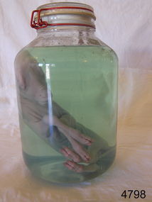
Animal specimen - Lamb Fetus
This lamb fetus has been placed in preserving fluid. This can be formaldehyde, isopropyl or ethanol. It is important that glass jars are used, as plastic will be affected by the chemicals over time in the preserving fluid. Note the glass lid and the rubber seal; also the plastic covered metal clasp that has no contact with the contents. It is not known how long the lamb has been in this jar, but it is remarkably well preserved with just a little film of scum on parts on the top of the interior of the jar.The use of such preserved specimens is widespread in teaching students of all ages, veterinary operatives and museums of the composition of certain animals, insects and birds. Any information about an animal — be it photographs, blood, feathers or fur samples — is better than no information at all. But specimens are vital to ground-truth.Large glass jar containing a lamb fetus in preserving fluid. Glass lid is secured with rubber seal and metal fastening. flagstaff hill, flagstaff hill maritime museum and village, warrnambool, maritime museum, maritime village, great ocean road, shipwreck coast, lamb, fetus, lamb fetus, animal specimen, biological specimen -
 Royal Australian and New Zealand College of Obstetricians & Gynaecologists (RANZCOG)
Obstetrical crotchet and blunt hook used by Box Hill Hospital labour ward
This is a destructive instrument which, when placed around the neck of the fetus and twisted, succeeded in severing the head. This type of instrument was used in emergency surgery when the fetus was already dead and the mother's condition was critical. This instrument might also have been used to remove tumors. Source: Yale University Library, 'Blunt hook and crochet', https://collections.library.yale.edu/catalog/17322011 This was included with other obstetric instruments, mostly destructive instruments, from Box Hill Hospital labour ward given to RANZCOG in February- March 1998. The maternity service at Box Hill hospital combined with St George's hospital in Kew to be known as Birralee Maternity Service. These instruments were collected by Julie Collette, Unit Manager, St George's Kew and given to RANZCOG Museum Curator, Susan Barnett.Obstetrical crotchet. Nickel plated stainless steel, with large blunt hook at one end and crotchet hook at the other, and fluting at the centre. "B.H.H.L. Ward"destructive instruments -
 Royal Australian and New Zealand College of Obstetricians & Gynaecologists (RANZCOG)
Tool - Cranioclast used by Dr Michael Kloss
A cranioclast was used to crush and extract the skull of a fetus to aid delivery in cases of difficult and obstructed labour. This instrument was part of a collection of instruments used by Dr Michael Kloss in his medical practice. Dr Kloss subsequently donated this collection to the College.Metal cranioclast. Instrument consists of two arms that join together with a pin. When joined, the arms wrap around each other for extra strength. Each arm has a handle for grip, and there is a ratchet with a wingnut at the end of the handles for widening the aperture between the arms and locking the arms in place. Each arm of the instrument is engraved with the word 'Kloss'.'Kloss'obstetrics, destructive instruments -
 Royal Australian and New Zealand College of Obstetricians & Gynaecologists (RANZCOG)
teaching model, female reproductive organs, 1940s-1950s
Originally this teaching model belonged to Prince Henry's hospit�l library and was transferred to the Monash Medical Centre, Clayton in the 1970s by Sister Gertrude Berger, a famous nurse-educator, who is best known for her work leading up to the transfer of nursing education in Victoria from hospitals to universities in 1986.Gerty (as she was known in the School of Nursing) bought them in Europe in the late 1940s and early 1950s.Model of female uterus with cross-section revealing a foetus, also, fallopian tubes and ovaries with cut away section revealing vagina and cervix. Painted plastic on metal rod with round wooden stand. Labelled on base "AV618.2 / 01 Fetus". Stamped on base, "PRINCE HENRY'S HOSPITAL/ NURSES LIBRARY." Label underneath base"Adam Rouilly/ London England".teaching model, anatomy, female uterus -
 Royal Australian and New Zealand College of Obstetricians & Gynaecologists (RANZCOG)
teaching model,foetus, 1940s-1950s
Originally this teaching model belonged to Prince Henry's hospit�l library and was transferred to the Monash Medical Centre, Clayton in the 1970s by Sister Gertrude Berger, a famous nurse-educator, who is best known for her work leading up to the transfer of nursing education in Victoria from hospitals to universities in 1986.Gerty (as she was known in the School of Nursing) bought them in Europe in the late 1940s and early 1950s.Model of a foetus, approximately seven weeks. Painted plastic on metal rod with round wooden stand. Labelled on base "AV618.2 / 01 Fetus/ FOETUS". Stamped on base, "PRINCE HENRY'S HOSPITAL/ NURSES LIBRARY." Label on base"Adam Rouilly/ London England". The model has anaaatomical numberings in black ink. There must have been an acompaning information booklet [missing].teaching model anatomy, female foetus -
 Royal Australian and New Zealand College of Obstetricians & Gynaecologists (RANZCOG)
Embryotomy scissors used by Box Hill Hospital labour ward, Thomas Russ & Son Ltd, Sheffield, England
Embryotomy is a destructive procedure whereby a fetus is dismembered to facilitate removal from the uterus when delivery is obstructed or impossible. It carries a risk of trauma (such as uterine rupture or vaginal/cervical injury) but aims to reduce the risks of mortality and impaired function to the patient. (Source: Medecins Sans Frontieres, 'MSF medical guidelines - Essential obstetric and newborn care - 9.7 Embryotomy'') This instrument was included with other obstetric instruments, mostly destructive instruments, given to RANZCOG from Box Hill Hospital labour ward in February- March 1998. The maternity service at Box Hill Hospital combined with St George's Hospital in Kew to be known as Birralee Maternity Service. These instruments were collected by Julie Collette, Unit Manager, St George's Kew and given to RANZCOG Museum Curator, Susan Barnett.Embriotomy scissors. Curved-on-flat scissors with take-off point.destructive instruments -
 Royal Australian and New Zealand College of Obstetricians & Gynaecologists (RANZCOG)
Simpson's cranioclast used by Dr Mitchell Henry O'Sullivan
Prior to the 1900s, complicated births, particularly where there was a disproportion between the size of the woman’s pelvis and the foetus’ head, often meant the death of the baby and the mother. Instruments for removing a dead or ailing foetus from within the mother were used to attempt to save the mother’s life. The cranioclast, first invented by Dr. James Simpson in the mid-19th century and later redesigned by others, was used for fetal destruction and removal. Fundamentally a strong pair of forceps, the cranioclast was used to crush the skull, decreasing its diameter. In some cases, this would allow normal uterine contractions to expel the foetus; in others, the physicians would use an obstetrical hook to pull the body out of the mother. Doctors disagreed as to the pelvic diameter that would necessitate this drastic intervention, but generally found that 3 to 3.5 inches was the smallest size through which a living infant could pass. Equally of debate was the pelvic size through which the dead fetus could be extracted. When vaginal extraction was deemed unadvisable, Caesarian section would be performed. As caesarean section became safer and more common with the advent of anaesthetics and antiseptic techniques, the use of cranioclasts and obstetrical hooks diminished. (Museum of Health Care, Kingston) Dr Mitchell Henry O'Sullivan worked in the Victorian country town of Casterton as a general practitioner from 1919 until his death in 1977. He also practiced obstetrics. His son, Dr David More O'Sullivan donated his obstetric bag and its contents to the College in 1999. The bag and contents are a unique time capsule of the type of instruments and pharmaceuticals used in the inter-war period.Hinged metal tool with bakelite handles at one end and serrated teeth at other end. The instrument is in two sections. The right or upper blade has a black bakelite handle. There are two screws on the inside of the handle, 5.5cm apart. In the centre of the blade is a screw notch in the shape of a small horseshoe. On the inner side of the blade is a depression extending most of the length. The left, or lower, blade also has a black bakelite handle. There are two screws on the inside of the handle approximately 6cm apart. Mobile metal clasps in the shape of an 'S' , with three serrations, is attached to the distal end of the handle, which enables the blades to be opened or closed. destructive instruments
