Showing 2 items matching "funduscopy"
-
 The Cyril Kett Optometry Museum
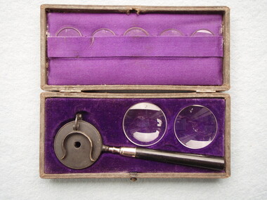
Equipment - Ophthalmoscope, unknown, Liebreich ophthalmoscope, cased, 1875 (estimated); late 19th century
Richard Liebreich of Germany invented his design of ophthalmoscope in 1855. This example is complete in its case with Coccius lenses and condensing lenses. Early ophthalmoscopes required an external source of illumination, eg lamp or candle, and light was reflected into the eye to be examined by the mirror on the ophthalmoscope. The earliest versions of the Liebreich ophthalmoscope used a polished metal surface to reflect light; glass mirrors were introduced in 1870. A condensing lens was held in front of the patient to view the image. A Coccius lens could be clipped into the holder to counter ametropia of user or subject.This Liebreich ophthalmoscope is significant for the collection as it is the only complete example of the three held in the collection.Cased Liebreich ophthalmoscope with 5 small coccius glass lenses and 2 glass condensing lenses. Non-illuminated ophthalmoscope has concave mirror in round head with central sight hole.Hinged coccius clip attached to hold lenses. Black metal head, silver coloured mount and black turned timber handle. Case has black leather outer lining and purple velvet and satin inner linings. Case hinged with snap closure. On front of case:"LIEBREICH'S OPHTHALMOSCOPE" 4 of 5 Coccius lenses engraved with powers: "8-", "12-", "-01", "+01"ophthalmoscope, optometry, ophthalmology, liebreich, coccius, lenses, eye examination, fundus, funduscopy, non illuminated, instrument, eye doctor, liebreich ophthalmoscope -
 Bendigo Historical Society Inc.
Instrument - Ophthalmoscope (or funduscope)
Ophthalmoscopy, also called funduscopy, is a test that allows a health professional to see inside the fundus of the eye and other structures using an ophthalmoscope (or funduscope). It is done as part of an eye examination and may be done as part of a routine physical examination. It is crucial in determining the health of the retina, optic disc, and vitreous humor. The pupil is a hole through which the eye's interior can be viewed. For better viewing, the pupil can be opened wider (dilated; mydriasis) before ophthalmoscopy using medicated eye drops (dilated fundus examination). However, undilated examination is more convenient (albeit not as comprehensive), and is the most common type in primary care. The Photoscope, or Mirror. Many varieties of the photoscope are in use. .... Some prefer a mirror with a small, folding, protecting handle, or two mirrors, so made that one may serve for the handle while the other is in use. These are easily carried in the pocket, ... If the mirror is perforated in the center, the light rays pass freely to the examiner's eye, but the edge of the perforation, unless perfectly blackened and free from chipping, causes very annoying reflections.Round circular black compact hinged - When opened has mirror with hole in it on one side and black lid/cover on the other - Small lip on top cover. ophthalmoscope, fundoscope, eye testing
