Showing 8 items matching "spectroscopy"
-
 University of Melbourne, School of Chemistry
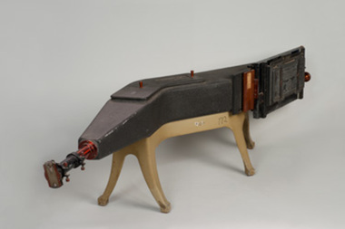
Spectogragh
Hilger Medium Quartz Spectrograph. Bought for use in early Raman Spectroscopy -
 The Ed Muirhead Physics Museum
Gas X-ray Tube
The investigation of the x-ray appears early on to have been a priority research topic at the University of Melbourne’s School of Physics. This interest was sparked by the appointment in 1889 of Professor T.R. Lyle. Lyle, who was head of the school until 1915, is thought to have been the first person in Australia to have taken an x-ray photograph. A copy of this photograph can be found in the School of Physics Archive. For this particular experiment Lyle actually made his own x-ray tube. His successor, Professor Laby, continued to work with x-rays. During the 1920s Laby worked on the x-ray spectra of atoms and in 1930 he co-published with Dr. C.E. Eddy, Quantitative Analysis by X-Ray Spectroscopy. Also with Eddy, Laby produced the landmark paper Sensitivity of Atomic Analysis by X-rays. Laby went on to have an x-ray spectrograph of his own design manufactured by Adam Hilger Ltd. (see cat. No. 38). School of Physics, the University of Melbourne Cat. No. 22. Jacqueline Eager Student Projects Placement, Cultural Collections 2005 The original X-ray tubes relied on low pressure operation. The electrons and positive ions are produced in the residual gas. Positive ions are accelerated towards the cathode and release electrons which on hitting the anode produce X-rays. These early gas X-ray tubes operated satisfactory only over a narrow pressure range. Stamped Label: “NATURAL PHILOSOPHY LABORATORY/ No/ UNIVERSITY OF MELBOURNE” Stamped: “90268 M. No. 5171[??]/No. 2156[??]/ M. No. 346585.” x-ray tubes, gas x-ray tube, laby, spectroscopy -
 The Ed Muirhead Physics Museum
Sight, between 1857 and 1948
Related to object # 467.1Cylindrical black sight with metallic fixtures. Adjustable base with screws.Side of barrel: 'E. R. WATTS & SON / LONDON / NO 19504' Under rear base mount: '2T'er watts & sons, sight, optical equipment, spectroscopy -
 The Ed Muirhead Physics Museum
Sight, between 1857 and 1948
Associated with object 467.2Cylindrical black sight with metallic fixtures and split sight. Adjustable base with screws.Logo in triangle on side of barrel: 'ERW&S' Stamped on underside of base: '2C'er watts & sons, spectroscopy, sight, optical equipment, surveying -
 Flagstaff Hill Maritime Museum and Village
Instrument - Marine Telescope, 1870-1880
This telescope was amongst various items collected from a sea dive in Port Phillip Bay. The diver was the caretaker of the Port Lonsdale Lighthouse, who dived on various wrecks in the bay during the 1960's. After the caretaker's death, his son sold off many of the shipwreck artefacts. The telescope was purchased from the caretaker's son in the 1990's by a previous owner of the Marine Shop, Queenscliff, Victoria. John Browning was particularly well known for his scientific advances in the fields of spectroscopy, astronomy, and optometry. Between 1856 and 1872, Browning acquired provisional patents for designs of numerous scientific instruments. He was also the recipient of an award at the 1862 International Exhibition held in London. Also recognised for his temperature-compensated aneroid barometer. Browning's scientific instruments were used in physics, chemistry, and biology. The products he designed and manufactured included spectroscopes, telescopes, microscopes, barometers, photometers, cameras, ophthalmologist, and electrical equipment such as electric lamps. John Browning was born around 1831 in Kent, England. His father, William Spencer Browning, was a maker of nautical instruments. John Browning's great-grandfather was also an instrument maker as well as John’s brother Samuel Browning of the firms Spencer & Browning and Spencer, Browning & Rust, who also manufactured navigational instruments. The latter firm was in operation in London from 1784 to 1840 and was succeeded by the firm of Spencer, Browning & Co. John Browning initially intended to follow the medical profession and entered Guy's Hospital, a teaching hospital and a school of medicine. Despite having passed the required examinations, however, he abandoned his plans. Instead, he apprenticed with his father, William Spencer Browning. At the same time, in the late 1840s, he was a student attending the Royal College of Chemistry several days per week. By the early 1870s, practical optics had become John Browning's primary interest, and he listed his occupation as an optician on the census records from 1871 to 1901. He was well known among London's ophthalmic surgeons for his various ophthalmic instruments. He had a large part in reforming the art of crafting spectacles. Other achievements were as an author of the book, How to Use Our Eyes and How to Preserve them by the Aid of Spectacles. Published in 1883, the book included thirty-seven illustrations, including a diagram demonstrating the anatomy of the eye. In 1895, he was one of the founders of the "British Ophthalmology" the first professional organisation for optometry. He was not only its first president but also registered as its first member so many considered him to be the first professional optometrist. Other professional organisations he belonged too was as a member of “The Aeronautical Society of Great Britain”. In 1871 constructing the first wind tunnel located at Greenwich Marine Engineering Works. He was also a member of other scientific organisations, such as the “Microscopical Society of London”, the “Meteorological Society”, and the “Royal”. Then in 1908 the company of W. Watson & Son, opticians and camera makers, took over John Browning's company since 1901 John Browning had been semi-retired but in 1908 he fully retired and moved to Bournemouth in Hampshire. He died in Cheltenham, Gloucestershire in 1925.The telescope is significant for its association with one of the world’s leading scientific instrument makers and inventor of the 19th and early 20th century. It is believed the donation came off a wreck either in Port Philip Bay or between Point Lonsdale and the Nepean Heads making it a significant maritime historical artefact. Its provenance is good given it was taken off a wreck in this area by the Point Lonsdale lighthouse caretaker. Examples of John Browning's telescopes because of their scientific and historical importance are highly valued by collectors.Marine style single draw brass telescope with a sunshade. The single draw has no split and the second cartridge is held in a long brass tube within the single draw, mounted from the objective end. The eyepiece is flat and at the end of the first draw in a very faded engraving that is believed to read "John Browning, 63 Strand, and should read London under the word strand but this is hard to establish given the engravings condition. This interpretation of the engraving has been arrived at by examination of other John Browning telescope engraving examples."John Browning, engraved to the first tube in copper plate style "63 STRAND" Engraved under in capital textflagstaff hill, flagstaff hill maritime museum and village, warrnambool, maritime museum, maritime village, great ocean road, shipwreck coast, shipwreck artefact, port phillip bay, port lonsdale lighthouse, wreck, 1960’s diver, queenscliff marine shop, john browning, telescope, spectroscopy, optometry, scientific instruments, william spencer browning, optician, navigational instrument, microscopical society of london, aeronautical society, marine technology -
 The Ed Muirhead Physics Museum
Rotating Anode X-ray Tube
The investigation of the x-ray appears early on to have been a priority research topic at the University of Melbourne’s School of Physics. This interest was sparked by the appointment in 1889 of Professor T.R. Lyle. Lyle, who was head of the school until 1915, is thought to have been the first person in Australia to have taken an x-ray photograph. A copy of this photograph can be found in the School of Physics Archive. For this particular experiment Lyle actually made his own x-ray tube. His successor, Professor Laby, continued to work with x-rays. During the 1920s Laby worked on the x-ray spectra of atoms and in 1930 he co-published with Dr. C.E. Eddy, Quantitative Analysis by X-Ray Spectroscopy. Also with Eddy, Laby produced the landmark paper Sensitivity of Atomic Analysis by X-rays. Laby went on to have an x-ray spectrograph of his own design manufactured by Adam Hilger Ltd. (see cat. No. 38). School of Physics, the University of Melbourne Cat. No. 22. Jacqueline Eager Student Projects Placement, Cultural Collections 2005 A modern X-ray tube differs little from the original Coolidge tube. A minor modification is the rotating anode type that extends the life and increases the available power of the tube by presenting a new portion of the anode when required. “P125/20/40/NrF038803 (?) SIEMENS-REINIGER-WERRE AG ERLANGEN Eigen filleung (?) mind. 0,7 mm AL” On rotating shaft: “FO/33803” On cathode: “23C” -
 The Ed Muirhead Physics Museum
Gas X-ray Tube, Victor
The investigation of the x-ray appears early on to have been a priority research topic at the University of Melbourne’s School of Physics. This interest was sparked by the appointment in 1889 of Professor T.R. Lyle. Lyle, who was head of the school until 1915, is thought to have been the first person in Australia to have taken an x-ray photograph. A copy of this photograph can be found in the School of Physics Archive. For this particular experiment Lyle actually made his own x-ray tube. His successor, Professor Laby, continued to work with x-rays. During the 1920s Laby worked on the x-ray spectra of atoms and in 1930 he co-published with Dr. C.E. Eddy, Quantitative Analysis by X-Ray Spectroscopy. Also with Eddy, Laby produced the landmark paper Sensitivity of Atomic Analysis by X-rays. Laby went on to have an x-ray spectrograph of his own design manufactured by Adam Hilger Ltd. (see cat. No. 38). School of Physics, the University of Melbourne Cat. No. 22. Jacqueline Eager Student Projects Placement, Cultural Collections 2005 The original X-ray tubes relied on low pressure operation. The electrons and positive ions are produced in the residual gas. Positive ions are accelerated towards the cathode and release electrons which on hitting the anode produce X-rays. These early gas X-ray tubes operated satisfactory only over a narrow pressure range. Manufacturer’s mark stamped: “PATENTED/ VICTOR/ TRADEMARK/ MADE IN BOSTON U.S.A./ TUNGSTEN” A white circular stamp, stamped near the manufacturer’s mark: “[illegible]TER WIGGH[illegible]” Stamped label: “NAT. PHIL. LAB./ No./ UNIV. OF MELB.” Inscription on the end face of the copper piece: “PAT. SEP 5’ 11 DEC. 30’13/ JUNE 23, 14 NOV. 30.15/ 43835” -
 The Ed Muirhead Physics Museum
Coolidge X-ray Tube
The investigation of the x-ray appears early on to have been a priority research topic at the University of Melbourne’s School of Physics. This interest was sparked by the appointment in 1889 of Professor T.R. Lyle. Lyle, who was head of the school until 1915, is thought to have been the first person in Australia to have taken an x-ray photograph. A copy of this photograph can be found in the School of Physics Archive. For this particular experiment Lyle actually made his own x-ray tube. His successor, Professor Laby, continued to work with x-rays. During the 1920s Laby worked on the x-ray spectra of atoms and in 1930 he co-published with Dr. C.E. Eddy, Quantitative Analysis by X-Ray Spectroscopy. Also with Eddy, Laby produced the landmark paper Sensitivity of Atomic Analysis by X-rays. Laby went on to have an x-ray spectrograph of his own design manufactured by Adam Hilger Ltd. (see cat. No. 38). School of Physics, the University of Melbourne Cat. No. 22. Jacqueline Eager Student Projects Placement, Cultural Collections 2005 In 1913 Coolidge overcame the limitation of the narrow operating range of the gas X-ray tubes with the invention of the vacuum X-ray tube. A filament heated by an electric current directly releases electrons by thermionic emission. In thermionic emission, electrons are emitted from a metal surface directly by the application of an electric current to heat a wire filament. The electrons accelerate to the anode and produce X-rays. The anode has associated cooling fins due to the high temperatures attained by the release of kinetic energy by the electrons on colliding with the anode. Internal Glass sleeve: “A941/L2593/2821”
