Showing 24 items matching "cannula"
-
 Royal Australian and New Zealand College of Obstetricians & Gynaecologists (RANZCOG)
Instrument - Cannula, Chimco
... Cannula...Plastic cannula ...Royal Australian and New Zealand College of Obstetricians & Gynaecologists (RANZCOG) 1 Bowen Crescent Naarm (Melbourne) melbourne Plastic cannula Instrument Cannula Chimco ...Plastic cannula -
 Geoffrey Kaye Museum of Anaesthetic History
Equipment - Cannula, Transfusion
... ...Cannula...A selection of metal cannula of various designs and sizes....Equipment Cannula, Transfusion ...Doctors trained in blood transfusion were essential to the development of Forward Resuscitation Teams during World War I. In 1918, Dr Alan Holmes a Court and his colleagues established a resuscitation team and, following their remarkable success at the battle of Hamel on July 4, teams were permanently established at each of the five Australian divisions. Each team consisted of one doctor trained in surgery, blood transfusion and resuscitation, another doctor trained in anaesthesia, resuscitation and blood donor classification, and four other assisting staff. This team moved out to the wounded, rather than waiting for them to be stretchered back. They provided on-the-spot, life-saving resuscitation. The wounded were then transported back to the Casualty Clearing Station or Regimental Aid Post for further treatment.A selection of metal cannula of various designs and sizes.blood, transfusion, intravenous, cannula -
 Geoffrey Kaye Museum of Anaesthetic History
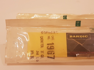
Equipment - Cannula placement set, Bardic, c. 1980
... ...Cannula...Clear plastic strip adhered to white paper backing, forming a sealed packet containing a cardboard backing board, with a cannula attached....Equipment Cannula placement set Bardic ...Cholera swept a deadly path through Europe in 1832. Irish physician, William O’Shaughnessy, proposed treating patients with saline infusions and Dr Thomas Latta of Leith, successfully applied the treatment. The intravenous route is the fastest way to deliver fluids and medications through the body. Today, fluid therapy is one of the most widespread interventions in acute medicine. Clear plastic strip adhered to white paper backing, forming a sealed packet containing a cardboard backing board, with a cannula attached.Stamped in black ink on 3929.1: CAT: / NO. 1966 / CATHETER: 14 GA. / .058 I.D. / 5 1/2 IN. / 0182037 Stamped in black ink on 3929.2: CAT: / NO. 1967 / CATHETER: 16 GA. .044 I.D. / 5 1/2 IN. / 0189037intravenous, cannula, fluid therapy, william o'shaughnessy, thomas latta -
 South West Healthcare
Ear Syringe, Medical Equipment, 20th Century
... 1 Metal cannister; 1 removable bulb cannula; 1 tapered cannula; 1 plunger...South West Healthcare Ryot Street Warrnambool great-ocean-road metal ear syringe 1 Metal cannister; 1 removable bulb cannula; 1 tapered cannula; 1 plunger Medical Equipment Ear Syringe ...1 Metal cannister; 1 removable bulb cannula; 1 tapered cannula; 1 plungermetal ear syringe -
 Flagstaff Hill Maritime Museum and Village
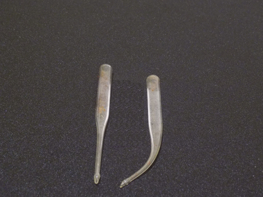
Trocar
... Cannula - outer sheath ...Cannula - outer sheath Trocar ...Trocar with Cannula, Trocar -metal 8 sided handle with sharp pointed end. Cannula - outer sheath flagstaff hill, warrnambool, shipwrecked-coast, flagstaff-hill, flagstaff-hill-maritime-museum, maritime-museum, shipwreck-coast, flagstaff-hill-maritime-village -
 Flagstaff Hill Maritime Museum and Village
Trocar
... Cannula - outer sheath ...Cannula - outer sheath Trocar ...Trocar with Cannula, Trocar -metal 4 sided grooved handle with sharp pointed end. Cannula - outer sheath flagstaff hill, warrnambool, shipwrecked-coast, flagstaff-hill, flagstaff-hill-maritime-museum, maritime-museum, shipwreck-coast, flagstaff-hill-maritime-village -
 South West Healthcare
Ear Irrigation Syringe, 20th century
... Chrome Metal Cannister; 1 tapered nozzle cannula and screw top plunger. ...South West Healthcare Ryot Street Warrnambool great-ocean-road Pomeroy Ear Irrigation Syringe: ear irrigation syringe syringe surgical instrument "British Make" 13 Chrome Metal Cannister; 1 tapered nozzle cannula and screw top plunger. Ear Irrigation Syringe British Make ...Pomeroy Ear Irrigation Syringe: Chrome Metal Cannister; 1 tapered nozzle cannula and screw top plunger. "British Make" 13ear irrigation syringe, syringe, surgical instrument -
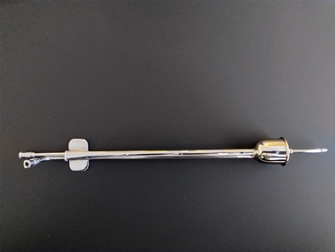
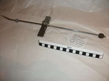 Royal Australian and New Zealand College of Obstetricians & Gynaecologists (RANZCOG)
Cannula, Spackman's, c1969
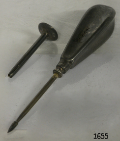
... This Spackman's cannula was used by Dr Geoff Bishop during gynaecological laparscopioc surgery.He used this decice as a uterine elevator. ...Manufacturers stamp: ANAX. Cannula, Spackman's ...Part of the laparoscopy equipment donated by Dr Geoff Bishop. Dr Geoffrey Bishop, whilst at the Department of O and G, University of Liverpool, UK, began laparoscopy in 1969. On returning to Australia, Bishop and Grimwade together with Mr Peter Paterson introduced gynaecological laparoscopy to Melbourne, practising at the Queen Victoria Memorial Hospital (QVMH), Melbourne in 1969. The College, through the Victorian State Committee of the Australian Council, RCOG, ran training courses in laparoscopy for local and interstate gynaecologists. These were conducted by Bishop, Grimwade and Paterson. They established protocols, with particular reference to safety, for the conduct of laparoscopy. Laparoscopy was used initially for diagnosis and for limited treatment using diathermy for conditions such as endometriosis. The real impetus came with the great upsurge of tubal sterilization in the early 1970s. Early techniques included diathermy and division of the Fallopian tubes using the Palmer forceps. [Dr Peter Renou, former honoury curator.]This Spackman's cannula was used by Dr Geoff Bishop during gynaecological laparscopioc surgery.He used this decice as a uterine elevator. Also, for testing tubal patency by inserting dye through it. Manufacturers stamp: ANAX.laparoscopy, tubal ligation, infertility investigation -
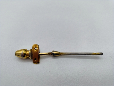 Royal Australian and New Zealand College of Obstetricians & Gynaecologists (RANZCOG)
Tool - Hamilton Bailey IV cannula used at Mackay Base Hospital, c. 1945-1960
... Gold coloured metal cannula. Consists of a hollow stem with eyelets on each of two sides, attached to a rectangular wedge-like plate with two holes through it. ...Tool Hamilton Bailey IV cannula used at Mackay Base Hospital ...Gold coloured metal cannula. Consists of a hollow stem with eyelets on each of two sides, attached to a rectangular wedge-like plate with two holes through it. This plate sits above a vase shaped fitting that attaches to a tube. -
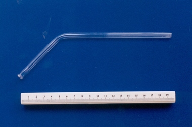 Royal Australian and New Zealand College of Obstetricians & Gynaecologists (RANZCOG)
Glass vaginal irrigator associated with midwife Mary Howlett, c. 1866 - 1920
... The glass tube is similar in appearance, however, to one of the four types of Brewer's glass cannula used in direct blood transfusion. (Reference Down Bros, page 958A)...The glass tube is similar in appearance, however, to one of the four types of Brewer's glass cannula used in direct blood transfusion. (Reference Down Bros, page 958A) Mary Howlett (1840-1922) began practising as a country midwife in 1866 in the western district of Victoria. ...The object has been identified as a vaginal (douche) irrigator. The glass tube is similar in appearance, however, to one of the four types of Brewer's glass cannula used in direct blood transfusion. (Reference Down Bros, page 958A)Mary Howlett (1840-1922) began practising as a country midwife in 1866 in the western district of Victoria. She qualified as a 'ladies monthly nurse' in 1887 and continued to practise as a nurse and midwife until 1920.She began her six months training at the Melbourne Lying-In Hospital. She was known by many as 'Auntie', and her career spanned more than 50 years. Mrs Howlett's midwifery box and contents were given to Dr Frank Forster, and he donated them to the museum collection in 1993.Glass tube, which functions as a vaginal irrigator. There is a curve in the tube, and it gets wider at proximal (far) end. The distal (near) end is round and blunt for attachment to rubber tubing.irrigation, midwifery -
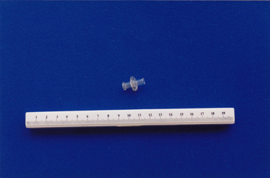
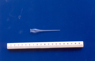 Royal Australian and New Zealand College of Obstetricians & Gynaecologists (RANZCOG)
Glass cannula associated with midwife Mary Howlett, c. 1866 - 1920
... Used to either irrigate the eye, instill medicated drops or tasks such as wound irrigation or the evacuation of fluid under the skin. Cannulas (or eye droppers as they are commonly called) were used both in homes and hospitals during the late 1880s and the early 1900s and were commonly available at chemists. ...Finely tapered at one end, with an open ended bulb at the other end. Glass cannula associated with midwife Mary Howlett, c. 1866 - 1920 ...Used to either irrigate the eye, instill medicated drops or tasks such as wound irrigation or the evacuation of fluid under the skin. Cannulas (or eye droppers as they are commonly called) were used both in homes and hospitals during the late 1880s and the early 1900s and were commonly available at chemists. The long tapered end gave the operator control over the rate of flow of the fluid in the bulb.Mary Howlett (1840-1922) began practising as a country midwife in 1866 in the western district of Victoria. She qualified as a 'ladies monthly nurse' in 1887 and continued to practise as a nurse and midwife until 1920.She began her six months training at the Melbourne Lying-In Hospital. She was known by many as 'Auntie', and her career spanned more than 50 years. Mrs Howlett's midwifery box and contents were given to Dr Frank Forster, and he donated them to the museum collection in 1993.Canula (or eye dropper) made of glass. Finely tapered at one end, with an open ended bulb at the other end. -
 Royal Australian and New Zealand College of Obstetricians & Gynaecologists (RANZCOG)
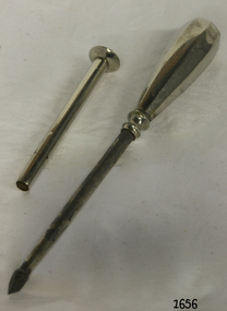
Infertility cannula used by Dr Geoff Bishop, Precious, c1969
... Cannula. Consists of a slender shaft with fittings at each end. ...Infertility Gynaecology Cannula. Consists of a slender shaft with fittings at each end. ...Part of a set of laparoscopy equipment donated by Dr Geoff Bishop. Dr Geoffrey Bishop, whilst at the Department of O and G, University of Liverpool, UK, began laparoscopy in 1969. On returning to Australia, Bishop and Grimwade together with Mr Peter Paterson introduced gynaecological laparoscopy to Melbourne, practising at the Queen Victoria Memorial Hospital (QVMH), Melbourne in 1969. The College, through the Victorian State Committee of the Australian Council, RCOG, ran training courses in laparoscopy for local and interstate gynaecologists. These were conducted by Bishop, Grimwade and Paterson. They established protocols, with particular reference to safety, for the conduct of laparoscopy. Laparoscopy was used initially for diagnosis and for limited treatment using diathermy for conditions such as endometriosis. The real impetus came with the great upsurge of tubal sterilization in the early 1970s. Early techniques included diathermy and division of the Fallopian tubes using the Palmer forceps. [Dr Peter Renou, former honorary curator.] This object was used by Dr Geoff Bishop during gynaecological laparoscopic surgery. This instrument is commonly used for suction. It is also used for testing tubal patency by inserting dye through it. Cannula. Consists of a slender shaft with fittings at each end. There are two points for tubal attachments at one end, set into separate small metal tubes at ninety degrees to each other. At the other end, there is a bell cap enclosing a curved nozzle. The nozzle is punctured with six small holes. Manufacturers stamp on plate near the attachment points reads 'PRECIOUS'.infertility, gynaecology -
 Royal Australian and New Zealand College of Obstetricians & Gynaecologists (RANZCOG)
Glass valve associated with midwife Mary Howlett, c. 1866 - 1920
... This valve connection may have attached to a syringe or cannula. Mary Howlett (1840-1922) began practising as a country midwife in 1866 in the western district of Victoria. ...Royal Australian and New Zealand College of Obstetricians & Gynaecologists (RANZCOG) 1 Bowen Crescent Naarm (Melbourne) melbourne This valve connection may have attached to a syringe or cannula. Mary Howlett (1840-1922) began practising as a country midwife in 1866 in the western district of Victoria. ...This valve connection may have attached to a syringe or cannula. Mary Howlett (1840-1922) began practising as a country midwife in 1866 in the western district of Victoria. She qualified as a 'ladies monthly nurse' in 1887 and continued to practise as a nurse and midwife until 1920. Mrs Howlett's midwifery box and contents were given to Dr Frank Forster and he donated to the museum collection in 1993. Small glass connection valve used for single flow. Body of valve consists of a hollow glass pipe with a lip at one end of the pipe and a central flange. intravenous device, midwifery -
 Geoffrey Kaye Museum of Anaesthetic History
Equipment - Cannulae, Transfusion
... ...Cannula...These transfusion needles were used to collect and administer blood for transfusions. Transfusion Cannula Kimpton Brown Blood blood transfusion Blundell Lower Denis Two glass tubes, one with straight and one with a curve at the base. ...Blood was long thought to be the essence of life and the centre of the soul; it was believed to provide a person with physical strength and mental abilities. In 1677, Richard Lower and Jean Baptiste Denis, in separate experiments, attempted animal-to-man transfusions to treat mental disorders. They had mixed success but didn't appear to cure the ailment. In 1818, James Blundell became interested in blood transfusion after witnessing the many deaths resulting from post-partum haemorrhage. He began with experiments in dogs and soon established it was possible to transfuse using a syringe if he worked quickly. Blundell established that cross-species transfusions didn't work and were dangerous. The early part of the 20th Century saw major developments in blood transfusion. Blood groups were identified by 1907 and the Kimpton Brown vessel (see 3675) slowed coagulation. These transfusion needles were used to collect and administer blood for transfusions.Two glass tubes, one with straight and one with a curve at the base. The tubes, known as cannualae, were used to facilitate blood transfusions.transfusion, cannula, kimpton brown, blood, blood transfusion, blundell, lower, denis -
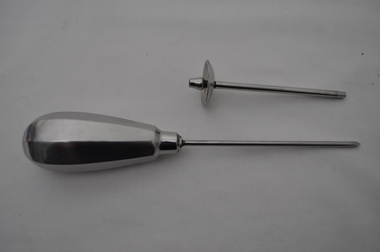
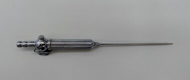 Royal Australian and New Zealand College of Obstetricians & Gynaecologists (RANZCOG)
Tool - Replica Veres needle, c. 2000
... Metal, spring loaded needle with an outer cannula and an inner stylus. Consists of a thin, hollow shaft with a bevelled point at one end, encasing a spring loaded needle/stylus with an eyelet at the end. ...Surgery Metal, spring loaded needle with an outer cannula and an inner stylus. Consists of a thin, hollow shaft with a bevelled point at one end, encasing a spring loaded needle/stylus with an eyelet at the end. ...This item was located in storage along with a newsletter from the Hungarian Society of Gynaeocological Endoscopists. The newsletter was produced in English to commemorate the ISGE (International Society for Gynaecological Endoscopy) meeting which was held in Hungary in August 2000. The newsletter contains an article on János Veres, the inventor of the needle, and an interview with his surviving family. It is possible that the replica needle was given as a gift to attendees of the meeting.Metal, spring loaded needle with an outer cannula and an inner stylus. Consists of a thin, hollow shaft with a bevelled point at one end, encasing a spring loaded needle/stylus with an eyelet at the end. Opposite end of needle consists of an open valve leading to the inner needle with a bulbous connection point. There is a round protrusion at the bottom of the bulbous connection point, and a small oval shaped grip at the top of this point, running parallel to the body of the instrument. A metal barrel containing the spring sits adjacent to the connection point. The barrel can be unscrewed to access the spring inside. surgery -
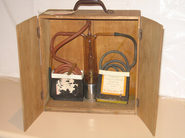 Royal Australian and New Zealand College of Obstetricians & Gynaecologists (RANZCOG)
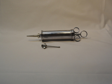
Rubin's tubal insufflator apparatus associated with St Vincent's Hospital, c1919
... It blows carbon dioxide, via a cannula, into the uterus. The ease with which gas escaped through the Fallopian tubes was reflected by pressure changes on an instrument called a manometer. ...It blows carbon dioxide, via a cannula, into the uterus. The ease with which gas escaped through the Fallopian tubes was reflected by pressure changes on an instrument called a manometer. ..."Potential blockage in the Fallopian tubes was assessed using this apparatus. It was developed by American gynaecologist Isidor Clinton Rubin (1883-1958). It blows carbon dioxide, via a cannula, into the uterus. The ease with which gas escaped through the Fallopian tubes was reflected by pressure changes on an instrument called a manometer. Blockage of the tubes is often due to previous infection or surgery. It is a common cause of infertility. Rubin’s test formed a standard part of infertility investigations for many years. It was gradually replaced by an X-ray technique involving radio-opaque ‘dye’ injected into the uterus." Source: Science Museum Group. Rubin’s apparatus for uterotubal insufflation, New York, United States, 1928. A639503Science Museum Group Collection Online. Accessed 12 June 2024. https://collection.sciencemuseumgroup.org.uk/objects/co96774/rubins-apparatus-for-uterotubal-insufflation-new-york-united-states-1928-tubal-insufflator. There is no manometer to monitor gas pressure on this model so it is either incomplete or a manometer was not available in this possibly early model. This device may be dated c1919, 1920s, or 1930s. 1919 was the year Isidor Clinton Rubin (1883-1958) introduced this apparatus. Rubin's tubal insufflator apparatus. Consists of a large cylindrical glass canister, with three glass nozzles at top with long rubber tubing attached to each. The device is inside a portable plywood box with two door. One surgical steel introducer, and one glass introducer, are also attached to the device. -
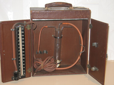 Royal Australian and New Zealand College of Obstetricians & Gynaecologists (RANZCOG)
Tubal insufflator associated with Dr Lorna Lloyd-Green, c1919
... It blows carbon dioxide, via a cannula, into the uterus. The ease with which gas escaped through the Fallopian tubes was reflected by pressure changes on an instrument called a manometer. ...It blows carbon dioxide, via a cannula, into the uterus. The ease with which gas escaped through the Fallopian tubes was reflected by pressure changes on an instrument called a manometer. ..."Potential blockage in the Fallopian tubes was assessed using this apparatus. It was developed by American gynaecologist Isidor Clinton Rubin (1883-1958). It blows carbon dioxide, via a cannula, into the uterus. The ease with which gas escaped through the Fallopian tubes was reflected by pressure changes on an instrument called a manometer. Blockage of the tubes is often due to previous infection or surgery. It is a common cause of infertility. Rubin’s test formed a standard part of infertility investigations for many years. It was gradually replaced by an X-ray technique involving radio-opaque ‘dye’ injected into the uterus." Source: Science Museum Group. Rubin’s apparatus for uterotubal insufflation, New York, United States, 1928. A639503Science Museum Group Collection Online. Accessed 12 June 2024. https://collection.sciencemuseumgroup.org.uk/objects/co96774/rubins-apparatus-for-uterotubal-insufflation-new-york-united-states-1928-tubal-insufflator. Model may be dated c1919 or 1920s or 1930s. 1919 was the year Isidor Clinton Rubin (1883-1958) introduced the apparatus. Instrumant has a label with Cyrus Jones monogram " Donated by Dr Lorna Lloyd Green, 1986/ Rubin's Insufflator/ NB "sparklet holder separate" missing?Rubin's tubal insufflator apparatus, large cylidrical glass canister inside a portable carry box with two doors with three glass nozzels at top with long rubber tubing attached on each. One surigical steel introducer, one glass introducer attached. A blood pressure manometer is fixed on the inside door. infertility -
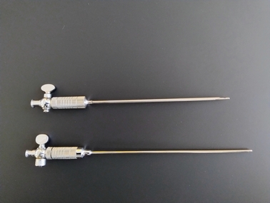 Royal Australian and New Zealand College of Obstetricians & Gynaecologists (RANZCOG)
Instrument - Two Veres needles used by Dr Geoff Bishop, WISAP Medical Technology GmbH
... Metal, spring loaded needle with an outer cannula and an inner stylus. Consists of a thin, hollow shaft with a bevelled point at one end, encasing a spring loaded needle/stylus with an eyelet at the end. ...Metal, spring loaded needle with an outer cannula and an inner stylus. Consists of a thin, hollow shaft with a bevelled point at one end, encasing a spring loaded needle/stylus with an eyelet at the end. ...Used for laparoscopy in gynaecology as well as in general surgery. Named for János Veres(s) (1903–1979), a Hungarian internist working with tuberculosis patients. At the time, one of the mainstays of treatment was to collapse an infected lung and allow lesions to heal. The needle was introduced as a safer technique to give patients such pneumothoraces. It was not until 1938, when he published his invention in the German literature, that the needle became more broadly known outside of Hungary. (Wikipedia) Believed to have been used and donated by Dr Geoff Bishop.Two Veres needles of differing lengths. Metal, spring loaded needle with an outer cannula and an inner stylus. Consists of a thin, hollow shaft with a bevelled point at one end, encasing a spring loaded needle/stylus with an eyelet at the end. Opposite end of needle consists of an open valve leading to the inner needle with a bulbous connection point. There is a round protrusion at the bottom of the bulbous connection point, and a small oval shaped grip at the top of this point, running parallel to the body of the instrument. A metal barrel containing the spring sits adjacent to the connection point. The barrel can be unscrewed to access the spring inside. The oval shaped grip on the shorter needle is engraved with the text 'WISAP/W. GERMANY'. The oval shaped grip on the longer needle is engraved with the number '3'. The number '30 is also engraved at the base of the barrel on the longer needle. 'WISAP/W.GERMANY', '3'gynaecology, surgery -
 Kiewa Valley Historical Society
Antral Trochar and Cannula
... Antral Trochar and Cannula ...This medical / hospital instrument was used in the Tawonga District General Hospital which was built in the 1950s specifically for the increase in population due to the Kiewa Hydro Scheme. This instrument was used in nasal surgery and is in a sterilised bag.Historically: Shows the development of scientific hospital equipment. Provenance: Used in the Tawonga District General Hospital which was remote and therefore required good equipment. Good condition and good interpretation capacity.In sterilised bag. Long pointed steel instrument with plastic handle with a protective shield a little along from the handle. The end of the steel rod has a very sharp point.medical instrument. hospital equipment. nasal. nose. mt beauty. tawonga. surgery., doctor. nurse -
 Ballarat Base Hospital Trained Nurses League
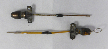
Venoclysis Cannula - Hamilton Bailey's
... Ballarat Base Hospital Trained Nurses League Drummond Street Nth Ballarat goldfields Venoclysis Cannula - Hamilton Bailey's Gold Plated Venoclysis Cannula - Hamilton Bailey's ...Gold Platedvenoclysis cannula - hamilton bailey's -
 Ballarat Base Hospital Trained Nurses League
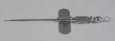
Trocar & Cannula for Intravenous Fluid Administration
... Ballarat Base Hospital Trained Nurses League Drummond Street Nth Ballarat goldfields Trocar & Cannula for Intravenous Fluid Administration Trocar & Cannula for Intravenous Fluid Administration ...trocar & cannula for intravenous fluid administration -
 Ballarat Base Hospital Trained Nurses League
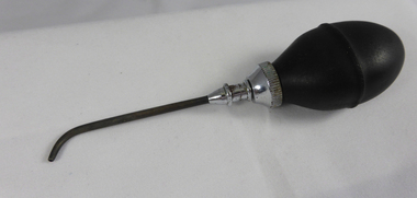
Ball Syringe with Cannula for Lavage
... Ballarat Base Hospital Trained Nurses League Drummond Street Nth Ballarat goldfields Ball Syringe with Cannula for Lavage Ball Syringe with Cannula for Lavage ...ball syringe with cannula for lavage -
 Ballarat Base Hospital Trained Nurses League
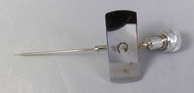
Metal Cannula
... Ballarat Base Hospital Trained Nurses League Drummond Street Nth Ballarat goldfields Metal Cannula Metal Cannula ...metal cannula -
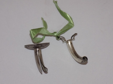 Geoffrey Kaye Museum of Anaesthetic History
Equipment - Cannula, Tracheostomy
... Attached to the square hook of the inner tube is a green cotton ribbon. Equipment Cannula, Tracheostomy ...This was gifted to Robin William Smallwood on his retirement. Smallwood graduated from medicine in the mid-1950s and decided to make a career in anaesthetics, was granted Fellowship in 1965, became a member of the Board of the Faculty in 1976 and became Dean in 1986-1987. It has been made by Arnold & Sons of London who were medical instrument manufacturers and became Mayer & Meltzer.Silver tube in two pieces, which form an innner and outer tube. The inner tube is curved with a flat plate at the top and two squared hooks (handles) coming off the plate. The outer tube has been spliced, creating two separate curved sides with an oval, bowl-like plate at the end, with an oval shaped holed punched through either side. Attached to the square hook of the inner tube is a green cotton ribbon.Stamped into the bowl shaped plate: ARNOLD & SONS / SILVERsmallwood, robin, •faculty dean, faculty of anaesthetists, royal australasian college of surgeons, ffaracs, racs, fanzca