Showing 27 items matching "urine"
-
 Moorabbin Air Museum
Container (Item) - Air Force Urine Collecting Device
... Air Force Urine Collecting Device....Air Force urine collecting device , with strap....Moorabbin Air Museum Moorabbin Airport 12 First Street Moorabbin melbourne Air Force urine collecting device , with strap. Air Force Urine Collecting Device. ... -
 Whitehorse Historical Society Inc.
Domestic object - Tes-Tape, urine sugar analysis tape, Eli Lily(Australia) & Co. Sydney, C 1970's
... Domestic urine sugar analysis paper...Silver cardboard box containing tape for domestic urine analysis Direction for use on box...Domestic urine sugar analysis paper Domestic medical aid Various, including detailed directions for use. ...Domestic urine sugar analysis paperSilver cardboard box containing tape for domestic urine analysis Direction for use on boxVarious, including detailed directions for use.domestic medical aid -
 Alfred Hospital Nurses League - Nursing History Collection
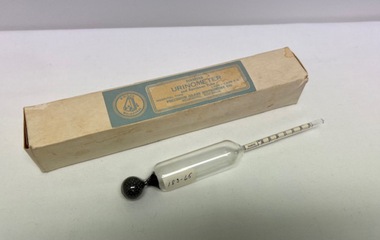
Instrument - Specific gravity urinometer, Diabetes Urinometer
... Glass urine specific gravity measure....Stem- Has calibrations with numbers marked to measure the specific gravity Missing urine holding specimen tube...Alfred Hospital Nurses League - Nursing History Collection Ground Floor, Building 10 Caulfield Hospital 260 Kooyong Road Caulfield melbourne Glass urine specific gravity measure. Urine specific gravity measure Urine testing Hospital type diabetes urinometer 60 degrees F Precision Glass Instrument Co. ...Glass urine specific gravity measure.Cream coloured manufacturers box labelled with a pale blue and cream label: "Diabetes Urinometer and Specimen Tube. Manufacturer Precision Glass Instrument Co, Melbourne". Blown glass urinometer used to measure the specific gravity of urine. It consists of 3 parts: I. The float: is the air containing part II. Weight: the lower end of urinometer (metal ball bearings) III. Stem- Has calibrations with numbers marked to measure the specific gravity Missing urine holding specimen tubeHospital type diabetes urinometer 60 degrees F Precision Glass Instrument Co. Melb. Measurements on stem from 000 to 030 urine specific gravity measure, urine testing -
 Dutch Australian Heritage Centre Victoria
Kruikezeiker (Jug Pisser) Statue
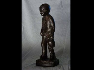
... A link to the folkloric custom that the poor sold their urine to the wool industry where it was used in wool washing. ...male figure depicting him about to fill a jug with his urine. His right hand lifts his garment while he holds the jug in his left. ...Dutch Australian Heritage Centre Victoria 60 Rosstown Road Carnegie melbourne A link to the folkloric custom that the poor sold their urine to the wool industry where it was used in wool washing. ...A link to the folkloric custom that the poor sold their urine to the wool industry where it was used in wool washing. Current depictions of a stereotypical Kruikezeiker are based on the Tilburg sculpture created by Henk Smulders (1925-1994). Said statue is located on Nieuwlandstraat, Tilburg. Tilburg has embraced this legend and named itself "Kruikezeiker Stad" (Jug City). The legend is brought to the fore during Karneval celebrations. It is a symbol of the city. Bronze (?) male figure depicting him about to fill a jug with his urine. His right hand lifts his garment while he holds the jug in his left. Base edge carries the name: "H.Smulders" on the back; "Tilburg" on the side and "Kruikezeiker" on the front.statue, tilburg, kruikezeiker -
![Equipment - Urinary receiver, Irving Urinary Receiver, [ca. 1930's]](/media/collectors/50187a8f023fd7201471f198/items/5a1c70a021ea670ba872884f/item-media/5a1c718d21ea670ba873490f/item-fit-380x285.jpg?cb=6) Alfred Hospital Nurses League - Nursing History Collection
Alfred Hospital Nurses League - Nursing History CollectionEquipment - Urinary receiver, Irving Urinary Receiver, [ca. 1930's]
... There were issues post-operatively as dressings were saturated with urine and causing excoriation. ...Supra pubic urinary drainage cover/receiver, metal with two drainage outlets to enable urine to drain via an abdominal drain tube following a supra pubic cystoscopy and prostatectomy, held in place by a leather belt, two rubber tubes were then attached other ends placed in a urinal...There were issues post-operatively as dressings were saturated with urine and causing excoriation. The receiver was said to be easy to apply, comfortable to wear and effective in keeping the patient dry and able to move freely in bed. ...This apparatus was devised by Hamilton Irving an English general surgeon in 1907. Supra pubic prostatectomy surgery was being performed more frequently due to improvements in procedures. There were issues post-operatively as dressings were saturated with urine and causing excoriation. The receiver was said to be easy to apply, comfortable to wear and effective in keeping the patient dry and able to move freely in bed. Supra pubic urinary drainage cover/receiver, metal with two drainage outlets to enable urine to drain via an abdominal drain tube following a supra pubic cystoscopy and prostatectomy, held in place by a leather belt, two rubber tubes were then attached other ends placed in a urinalFront of appliance has 'RAMSAY' inscribed as well as 'Wd 3' [Ward 3].post operative prostatectomy 1930s, medical equipment -
![Equipment - Urinary receiver, Irving Urinary Receiver, [ca. 1930's]](/media/collectors/50187a8f023fd7201471f198/items/673be1a8d073ef374cc00591/item-media/6743ce4fa758bc6936137eee/item-fit-380x285.jpg?cb=6) Alfred Hospital Nurses League - Nursing History Collection
Alfred Hospital Nurses League - Nursing History CollectionEquipment - Urinary receiver, Irving Urinary Receiver, [ca. 1930's]
... There were issues post-operatively as dressings were saturated with urine and causing excoriation. ...Supra pubic urinary drainage cover/receiver, metal with two drainage outlets to enable urine to drain via an abdominal drain tube following a supra pubic cystoscopy and prostatectomy, held in place by a leather belt, two rubber tubes were then attached other ends placed in a urinal...There were issues post-operatively as dressings were saturated with urine and causing excoriation. The receiver was said to be easy to apply, comfortable to wear and effective in keeping the patient dry and able to move freely in bed. ...This apparatus was devised by Hamilton Irving an English general surgeon in 1907. Supra pubic prostatectomy surgery was being performed more frequently due to improvements in procedures. There were issues post-operatively as dressings were saturated with urine and causing excoriation. The receiver was said to be easy to apply, comfortable to wear and effective in keeping the patient dry and able to move freely in bed. Supra pubic urinary drainage cover/receiver, metal with two drainage outlets to enable urine to drain via an abdominal drain tube following a supra pubic cystoscopy and prostatectomy, held in place by a leather belt, two rubber tubes were then attached other ends placed in a urinal"Ward 3" etched on to top and underneath rim. Old catalogued number "176-78" in black ink on underneath rim and sticker with "176-78" on side.post operative prostatectomy 1930s, medical equipment -
 Northern District School of Nursing. Managed by Bendigo Historical Society Inc.
Tool - Northern District School of Nursing Graduates Association - Urine and Faeces Testing Chart, 1950s
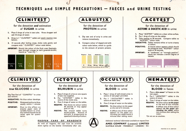
... Northern District School of Nursing Graduates Association - Urine and Faeces Testing Chart - printed in Great Britain by AMES Company (London) Limited This item contains the following document: 3792.10a Northern District School of Nursing Graduates Association - Urine and Faeces Testing Chart...Nurse Training Lister House history ames company pty ltd london Northern District School of Nursing Graduates Association - Urine and Faeces Testing Chart - printed in Great Britain by AMES Company (London) Limited This item contains the following document: 3792.10a Northern District School of Nursing Graduates Association - Urine and Faeces Testing Chart Tool Northern District School of Nursing Graduates Association - Urine and Faeces Testing Chart ...The Northern District School of Nursing in Victoria, Australia Graduates Association: History of the School Managed by a committee including hospital administrators, medical professionals, and nursing leaders from several regional hospitals. Associated with major hospitals in the Northern District, including Bendigo Base, Mildura Base, Castlemaine, Echuca, Swan Hill, St. Arnaud, and Kyneton District Hospitals. Provide high-quality theoretical and practical nursing training. Raise the standard of nursing education in the region. Maintain the highest principles of nursing practice. Applicants must be at least 17 years old and hold a Proficiency Certificate or higher educational qualification. Application involves submitting forms, certificates, an interview, an aptitude test, and a medical examination. A three-month probationary period follows initial acceptance. Preliminary Training School: Four-week introductory course covering basic nursing, anatomy, hygiene, and practical skills. Practical experience in various hospital departments, guided by senior staff. Study Block Plan: Includes several blocks of theoretical and practical instruction throughout the three-year program. Regular school exams and two state exams (First Professional and Final State) are required for progression and graduation. Nurses work a 40-hour week with at least one or two days off per week and three weeks of annual leave. Accommodation is provided in comfortable hostels with good facilities. Uniforms are supplied free; nurses provide their own shoes and stockings. Salaries and allowances are regulated, with deductions for board and free medical care. Additional Information Post-Graduate Opportunities. Senior positions require further experience and additional certificates (e.g., Midwifery, Infant Welfare).Diploma courses available in administration, teaching, and specialized nursing fields. Bursaries are available for advanced study.Career Prospects: Graduates can pursue roles in administration, teaching, ward and departmental leadership, district and visiting nursing, industrial and school nursing, and more. Opportunities existed both within hospitals and in community or specialized settings.FAQs and Practical Details. Minimal training costs (mainly exam fees and personal items).Living out allowances and travel expenses are covered. Four training intakes per year; waiting periods are short if qualifications are met.Northern District School of Nursing Graduates Association - Urine and Faeces Testing Chart - printed in Great Britain by AMES Company (London) Limited This item contains the following document: 3792.10a Northern District School of Nursing Graduates Association - Urine and Faeces Testing Chartnurse training, lister house, history, ames company pty ltd london -
 Royal Australian and New Zealand College of Obstetricians & Gynaecologists (RANZCOG)
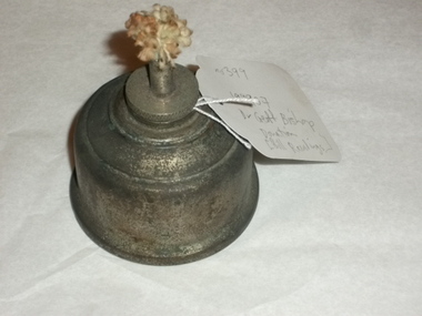
Spirit burner, 1940s
... Used by Bill Rawlings in process of testing urine....Used by Bill Rawlings in process of testing urine. Spirit burner ...This belonged to Dr Bill Rawling's and was in a medical bag that he was used in the 1940s and 1950s. It was donated by Dr Geoff Bishop.Spirit burner, metal container with thick woven cotton wick. No fuel present in container. Used by Bill Rawlings in process of testing urine.urine testing -
 Alfred Hospital Nurses League - Nursing History Collection
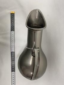
Functional object - monel metal female urinal, K.G.Luke
... Silver coloured metal female urine collector, funnel shaped neck with rolled metal opening, bulbous collecting chamber with flat base, curved handle to upper surface...Alfred Hospital Nurses League - Nursing History Collection Ground Floor, Building 10 Caulfield Hospital 260 Kooyong Road Caulfield melbourne used by nursing staff to assist women to void when bed ridden an essential item in providing care to non ambulant women monel metal female urinal K.G.Luke nursing care Alfred Hospital Alfred Hospital Nurses League Ward 4 176-024 black texta on base and sticky label blue ink to base, engraved - PARAMOUNT/STAINLESS STEEL/K.G.LUKE/63, WARD 4/AH engraved to superior surface Silver coloured metal female urine collector, funnel shaped neck with rolled metal opening, bulbous collecting chamber with flat base, curved handle to upper surface Functional object monel metal female urinal K.G.Luke ...used by nursing staff to assist women to void when bed riddenan essential item in providing care to non ambulant womenSilver coloured metal female urine collector, funnel shaped neck with rolled metal opening, bulbous collecting chamber with flat base, curved handle to upper surface176-024 black texta on base and sticky label blue ink to base, engraved - PARAMOUNT/STAINLESS STEEL/K.G.LUKE/63, WARD 4/AH engraved to superior surfacemonel metal, female urinal, k.g.luke, nursing care, alfred hospital, alfred hospital nurses league, ward 4 -
 Whitehorse Historical Society Inc.
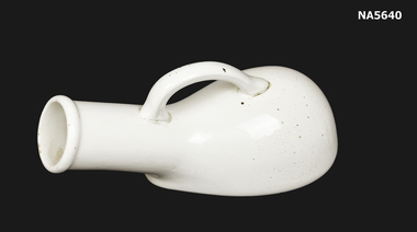
Equipment - Urinal, Unknown
... Used by male - urine relief hospital, home, portable...Whitehorse Historical Society Inc. 2-10 Deep Creek Road Mitcham melbourne Used by male - urine relief hospital, home, portable Hospital Medical Equipment For male use Fowler Ware Limited - Australia - Male use - Urinal bottle - porcelain - Fowler Ware - Handle Equipment Urinal Fowler Ware ...Used by male - urine relief hospital, home, portable- Male use - Urinal bottle - porcelain - Fowler Ware - Handle Fowler Ware Limited - Australiahospital, medical equipment, for male use -
 Alfred Hospital Nurses League - Nursing History Collection
Functional object - Urinary receiver for males, Urinal
... Urine...Donated by Mrs Bade Evans (nee Caldecott) Urine Urinal Enamel Mede in Czechoslovakia 24 White enamel urinal with handle and rim coloured blue Urinal Functional object Urinary receiver for males ...Donated by Mrs Bade Evans (nee Caldecott)White enamel urinal with handle and rim coloured blueMede in Czechoslovakia 24urine, urinal, enamel -
Northern District School of Nursing. Managed by Bendigo Historical Society Inc.
Tool - Northern District School of Nursing Graduates Association - Combistik Urine Test strips, 1950s
... Northern District School of Nursing Graduates Association - Combistik Urine Test strips - instruction booklet - 4 pages by the Ames Compant 65 Queens Road Melbourne This item contains the following document: 3792.11a Northern District School of Nursing Graduates Association - Combistik Urine Test strips - coloured instruction booklet Here are the key points from the document on Combistix urine test strips: Clinical Importance of UrinalysisUrinalysis is often underestimated but provides valuable diagnostic information.Simple to perform, yet highly informative for detecting urinary and metabolic conditions.Combistix is a combination test strip that rapidly screens for glucose, protein, and pH in urine. ...Tool Northern District School of Nursing Graduates Association - Combistik Urine Test strips ...The Northern District School of Nursing in Victoria, Australia Graduates Association: History of the School Managed by a committee including hospital administrators, medical professionals, and nursing leaders from several regional hospitals. Associated with major hospitals in the Northern District, including Bendigo Base, Mildura Base, Castlemaine, Echuca, Swan Hill, St. Arnaud, and Kyneton District Hospitals. Provide high-quality theoretical and practical nursing training. Raise the standard of nursing education in the region. Maintain the highest principles of nursing practice. Applicants must be at least 17 years old and hold a Proficiency Certificate or higher educational qualification. Application involves submitting forms, certificates, an interview, an aptitude test, and a medical examination. A three-month probationary period follows initial acceptance. Preliminary Training School: Four-week introductory course covering basic nursing, anatomy, hygiene, and practical skills. Practical experience in various hospital departments, guided by senior staff. Study Block Plan: Includes several blocks of theoretical and practical instruction throughout the three-year program. Regular school exams and two state exams (First Professional and Final State) are required for progression and graduation. Nurses work a 40-hour week with at least one or two days off per week and three weeks of annual leave. Accommodation is provided in comfortable hostels with good facilities. Uniforms are supplied free; nurses provide their own shoes and stockings. Salaries and allowances are regulated, with deductions for board and free medical care. Additional Information Post-Graduate Opportunities. Senior positions require further experience and additional certificates (e.g., Midwifery, Infant Welfare).Diploma courses available in administration, teaching, and specialized nursing fields. Bursaries are available for advanced study.Career Prospects: Graduates can pursue roles in administration, teaching, ward and departmental leadership, district and visiting nursing, industrial and school nursing, and more. Opportunities existed both within hospitals and in community or specialized settings.FAQs and Practical Details. Minimal training costs (mainly exam fees and personal items).Living out allowances and travel expenses are covered. Four training intakes per year; waiting periods are short if qualifications are met.Northern District School of Nursing Graduates Association - Combistik Urine Test strips - instruction booklet - 4 pages by the Ames Compant 65 Queens Road Melbourne This item contains the following document: 3792.11a Northern District School of Nursing Graduates Association - Combistik Urine Test strips - coloured instruction booklet Here are the key points from the document on Combistix urine test strips: Clinical Importance of UrinalysisUrinalysis is often underestimated but provides valuable diagnostic information.Simple to perform, yet highly informative for detecting urinary and metabolic conditions.Combistix is a combination test strip that rapidly screens for glucose, protein, and pH in urine. The test requires only one dip and 10 seconds to deliver three results, with no need for urine pre-treatment. Suitable for routine hospital urinalysis and large-scale screening, even by non-specialists due to its simplicity. Helps optimize laboratory workflow by allowing ancillary staff to perform basic tests, freeing technologists for more complex tasks. Useful for diagnosing and managing urinary tract infections, metabolic conditions, and monitoring therapy (e.g., sulphonamide therapy, chemotherapy for genito-urinary infections). Glucose: Essential for diabetes screening and management; Combistix is specific for glucose and does not react to lactose, making it reliable during pregnancy. Proteinuria is a key indicator of renal abnormalities, vascular disease, or pre-eclampsia. Its absence is reassuring. The test is sensitive and can be performed accurately by inexperienced users. The strip has three color-coded, reagent-impregnated areas separated by barriers for clear results.Simple procedure: dip, wait 10 seconds, and compare colors to a chart. Packaged in air-tight bottles of 100 strips, available through standard medical supply channels. Combistix offers a fast, reliable, and easy-to-use method for routine urine screening, supporting both clinical diagnosis and efficient laboratory operations.nurse training, lister house, ames company, combistix -
 Royal Australian and New Zealand College of Obstetricians & Gynaecologists (RANZCOG)
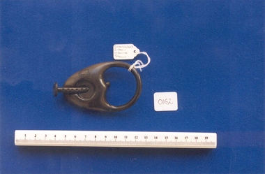
Greenhalgh stem pessary associated with Dr Frank Forster
... This ellipse shaped pessary, worn anteriorly, prevented dragging on the posterior wall of the bladder, thus preventing incontinence of urine often experienced by the wearing of a larger pessary....Royal Australian and New Zealand College of Obstetricians & Gynaecologists (RANZCOG) 1 Bowen Crescent Naarm (Melbourne) melbourne This ellipse shaped pessary, worn anteriorly, prevented dragging on the posterior wall of the bladder, thus preventing incontinence of urine often experienced by the wearing of a larger pessary. ...This ellipse shaped pessary, worn anteriorly, prevented dragging on the posterior wall of the bladder, thus preventing incontinence of urine often experienced by the wearing of a larger pessary.Vulcanite pessary. Pessary consists of a loosely tear shaped flange and a stem. Upper part of pessary is enclosed, with a keyhole opening for the stem. Upper part of body tapers to a stem. Lower part of body has been cut away, leaving a heart shaped opening. The stem is attached to the body through the keyhole opening. There are multiple perforations along the stem. intrauterine device, pessary -
 Royal Australian and New Zealand College of Obstetricians & Gynaecologists (RANZCOG)
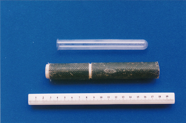
Test tube and case associated with midwife Mary Howlett, c. 1866 - 1920
... IT may also have been used to boil urine to identify the presence of urinary abnormalities such as sugar albumen acetone or bile....IT may also have been used to boil urine to identify the presence of urinary abnormalities such as sugar albumen acetone or bile. ...This type of test tube would have been used to collect blood or other bodily fluids. IT may also have been used to boil urine to identify the presence of urinary abnormalities such as sugar albumen acetone or bile.Mary Howlett (1840-1922) began practising as a country midwife in 1866 in the western district of Victoria. She qualified as a 'ladies monthly nurse' in 1887 and continued to practise as a nurse and midwife until 1920.She began her six months training at the Melbourne Lying-In Hospital. She was known by many as 'Auntie', and her career spanned more than 50 years. Mrs Howlett's midwifery box and contents were given to Dr Frank Forster, and he donated them to the museum collection in 1993.Glass test tube with a thin glass lip in original cylinder cardboard case. Case is in two section, lid and body - lid is lined with white cardboard.diagnostic testing, midwifery -
 Emerald Museum & Nobelius Heritage Park
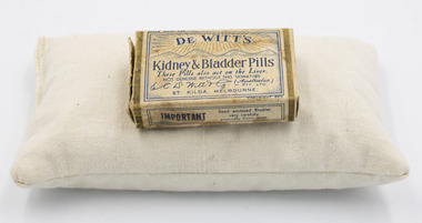
Container - Medicinal tablet box, unknown
... After taking these pills the urine will become a bluish or greenish colour. ...After taking these pills the urine will become a bluish or greenish colour. ...Off the shelf medication produced by a well known medications firm, who still exist today - De Witt.The pills and the associated instruction sheet give a view of how common ailments were dealt with in a previous era.A small pill box containing De Witts kidney & Bladder pills (3) & InstuctionsDe Witt's Kidney & Bladder Pills/These pills also act on the liver/Not genuine without this signature (Signed - E.C.De Witt)/Australia Pty. Ltd./St.kilda, Melbourne/De Witt's Kidney & Bladder pills - for weak kidneys, rheumatism, inflammation of the bladder, bachache, scalding or scanty urine, too frequent desire to urinate, gravel and all uric acid complaints. Cleanse the system. Act surely but gently on the liver/Dose - Adults take 1 pill before each meal and 2 at bedtime, with glassful of pure water. After taking these pills the urine will become a bluish or greenish colour. Do not be alarmed. Read enclosed booklet/enclosed lengthy instruction sheet -
St Vincent's Hospital Melbourne Archives
Book, Modern Practical Nursing Procedures, sixth edition by Doherty, Sirl and Ring in 1954
... There are extensive notes, some handwritten during lectures, for example on voluntary micturition there is an eleven- step procedure on how to encourage the patient to pass urine. This indicates the detailed learning nurses undertook whilst working at the same time in the wards....There are extensive notes, some handwritten during lectures, for example on voluntary micturition there is an eleven- step procedure on how to encourage the patient to pass urine. This indicates the detailed learning nurses undertook whilst working at the same time in the wards. ...One of the reference books used by Nurse Anne Carolan during her nurse training at St Vincents Hospital Melbourne in 1957. There are extensive notes, some handwritten during lectures, for example on voluntary micturition there is an eleven- step procedure on how to encourage the patient to pass urine. This indicates the detailed learning nurses undertook whilst working at the same time in the wards.Hand -written in ink and underlined inside the front cover is A Carolan's name. There are study notes written in pencil alongside the text on some pages. doherty sirl and ring, st vincent's hospital melbourne, nurse training, anne carolan, nursing procedures -
 City of Moorabbin Historical Society (Operating the Box Cottage Museum)
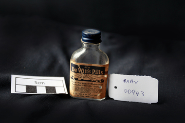
Manufactured Glass, bottle of 'De Witts Pills', mid 20thC
... It is not known how effective the pills were but the label assures people not to be alarmed if their urine takes on a bluish colour after taking a dose. ...It is not known how effective the pills were but the label assures people not to be alarmed if their urine takes on a bluish colour after taking a dose. ...De Witt's Kidney and Bladder Pills were produced by a firm founded in 1912 by E C De Witt, Cheshire UK, with branches in New Zealand, Chicago and New York. It became part of the CB Fleet Group in 1990, whose UK operation is currently based in Runcorn, Cheshire where they still manufacture toiletries, skin care products and pharmaceutical products. Aimed at adults and children above the age of eight, De Witt’s Kidney and Bladder Pills were intended to ease backache, aches and pains in the muscles, and kidney problems. They are typical of a huge range of treatments that were available ‘over the counter’ at pharmacists for many years. It is not known how effective the pills were but the label assures people not to be alarmed if their urine takes on a bluish colour after taking a dose. The indications or uses for this product as provided by the manufacturer are: A diuretic stimulant for the kidneys to promote the flow of urine, combined with analgesic action. Aids in alleviating muscular aches and pains, restlessness, dizziness, backache, headache, getting up at nights, loss of energy if caused by sluggish kidneys. An empty clear glass bottle with a metal screw top lid that contained De Witts Pills Front Label : NEW / DE WITT'S PILLS / rising sun trade mark / Relieve the pain of Rheumatism / Backache, Fibrositis, Sciatica / DIRECTIONS ......../ R.C.De Witt & Co (Aust) Pty Ltd. / St. Kilda Melbournepharmacy, medicines, glassware, bottles, moorabbin, bentleigh, cheltenham, de witt company ltd, cheshire england, cb fleet group ltd, glass manufacturers -
 Flagstaff Hill Maritime Museum and Village
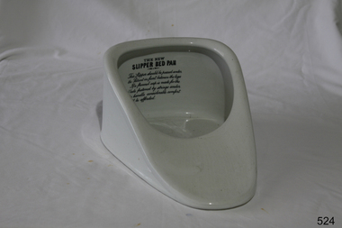
Equipment - Bed Pan
... The device is placed under the person's bottom, and it has a container that collects urine or stool. It is easy to clean and can be emptied into a toilet or sink. ...The device is placed under the person's bottom, and it has a container that collects urine or stool. It is easy to clean and can be emptied into a toilet or sink. ...The Bedpan Toilet device is a special tool designed for people who are unable to get out of bed to use the bathroom. It helps them go to the toilet comfortably and conveniently without having to leave their bed. The device is placed under the person's bottom, and it has a container that collects urine or stool. It is easy to clean and can be emptied into a toilet or sink. The Bedpan Toilet device allows individuals who are confined to bed due to illness or injury to maintain their dignity and independence by providing them with a practical solution for using the bathroom while staying in bed. The word bedpan was first seen in the literature of John Higgins in 1572, and one of the oldest known bedpans is on display in the Science Museum of London. It is a green, glazed earthenware bedpan that has been dated to the 16th or 17th century. At that time, bedpans were made from materials including pewter, brass, pottery, glass, and porcelain. Bedpans were not a commonplace item in hospitals until the late 1800s. Florence Nightingale, who worked as a nurse in the United Kingdom from the mid to late 1800s, recorded death rates and causes for soldiers in military hospitals during the Crimean War and then correlated them to corresponding sanitisation procedures. As a result, Nightingale proposed several methods to improve the sanitary conditions in both military and civilian hospitals, including the addition of bedpans in order to reduce infection exposure from urine or faeces. https://www.wikiwand.com/en/Bedpan The use of bedpans is significant, as it allows a patient who cannot move much, to remain in bed and perform toilet functions.Bed pan ceramic white glaze with handle. Labelled "The New Slipper Bed Pan". Has specific instructions for use under the maker's label.‘THE NEW SLIPPER BED PAN. This slipper should be passed under the patient in front between the legs. If a flannel cap is made for the blade fastened by strings under the handle considerable comfort will be afforded.’ flagstaff hill, warrnambool, shipwrecked-coast, flagstaff-hill, flagstaff-hill-maritime-museum, maritime-museum, shipwreck-coast, flagstaff-hill-maritime-village, nursing, bedpans, hygiene -
St Vincent's Hospital Melbourne Archives
Work on paper - Nursing training study notes belonging to Freda Fatzeus at St Vincent's Hospital Melbourne, 1925
... Each disease is described in length, how to nurse, what complications may occur and precautions to take, eg with Typhoid Fever, urine needs to be boiled to prevent spread of disease, container is to stand in antiseptic, kept separate, and if in the country, buried. ...St Vincent's Hospital Melbourne Archives Devonshire Arms building 38 Fitzroy Street Fitzroy melbourne Each disease is described in length, how to nurse, what complications may occur and precautions to take, eg with Typhoid Fever, urine needs to be boiled to prevent spread of disease, container is to stand in antiseptic, kept separate, and if in the country, buried. ...Each disease is described in length, how to nurse, what complications may occur and precautions to take, eg with Typhoid Fever, urine needs to be boiled to prevent spread of disease, container is to stand in antiseptic, kept separate, and if in the country, buried. Notes state:" the nurse must do all in her power to induce rest and sleep with the patients, eg warm sponge, frequent comfort positioning in bed." Other diseases detailed are Meningitis, Phthisis, Diptheria, Scarlet Fever, Measles, Pneumonia, all needing isolation nursing. How to prepare solutions give enemas and bowel washouts are described in these comprehensive notes indicative of the heavy, onerous nursing requirements practiced at that time. st vincent's hospital melbourne, nurse training, clinical notes, infectious diseases, freda fatzeus -
 Geoffrey Kaye Museum of Anaesthetic History
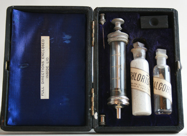
Equipment - Kit, Snake bite, Felton Grimwade & Co
... Treatments such as ammonia, strychnine, chlorinated lime, potassium permanganate, suction caps, alcohol, gunpowder, petrol, toad urine, iodide swabs and pig face plant juice were some that were used and sold. ...Treatments such as ammonia, strychnine, chlorinated lime, potassium permanganate, suction caps, alcohol, gunpowder, petrol, toad urine, iodide swabs and pig face plant juice were some that were used and sold. ...Prior to the first antivenom development in Australia, many of the snake men had a vast array of snakebite remedies they either used for themselves in the case of bites or pedaled to the public. Treatments such as ammonia, strychnine, chlorinated lime, potassium permanganate, suction caps, alcohol, gunpowder, petrol, toad urine, iodide swabs and pig face plant juice were some that were used and sold. The first antivenom produced in Australia was in 1930 for tiger snake bites. Subsequently, in response to public pressure, other antivenoms were produced. Taipan, 1955; Brown snake, 1956; Death Adder, 1958; Papuan black snake, 1959; Sea snake, 1961; and the polyvalent, 1962.Black box with hinged opening and gold leaf printed text on the top. Inside the box is lined with blue satin and velvet, and contains one (1) syringe with glass chamber and metal plunger and black rubber stopper, one (1) glass bottle with a label stating it contains chloride of lime, one (1) glass bottle with a label stating it contains pure alcohol and two (2) needles with metal connectors, one of which is broken. There is also a small wooden block with a groove in the top of it.Gold lettering on top of box: CHLORIDE OF LIME ANTIDOTE / FOR / SNAKE BITE / FELTON GRIMWADE & CO. / MELBOURNE.hydrochloride lime, alcohol, antidote, venom, snake bite -
 Royal Australian and New Zealand College of Obstetricians & Gynaecologists (RANZCOG)
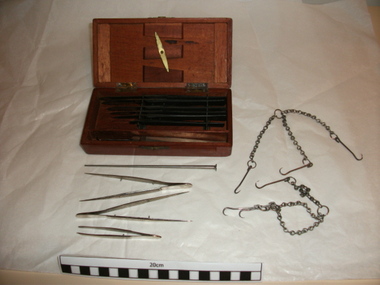
Surgical kit used by Lord Joseph Lister, Archibald Young of Edinburgh, 1870s
... Also included separately are two sets of autopsy hooks (hooks on chains), one metal blowpipe [commonly used with urine testing apparatus] and three sets of dissector forceps. ...Also included separately are two sets of autopsy hooks (hooks on chains), one metal blowpipe [commonly used with urine testing apparatus] and three sets of dissector forceps. ...This surgical instrument kit, c1870s, originally belonged to Lord Joseph Lister. On his retirement in 1892, Lord Lister presented the instrument kit to his friend Dr Alexander Matthew. The donor of the surgical kit, Professor Ian Stewart Fraser, is the grandson of Dr Alexander Matthew. The donor, Ian Fraser, checked with his mother about the inscription "Ethel Livie". There was no one of that name in his mother's family tree and the instruments were passed down from his mother's family. The inscription is faint and difficult to make out, and may also read 'Ethel Lavie' or 'Ethel Lane'.This surgical kit, made by Young of Edinburgh Scotland in the 1870s is significant because it belonged to and was most likely used by an internationally important figure in modern medicine, Lord Joseph Lister. Joseph Lister, 1st Baron Lister, Bt., OM, FRS, PC (5 April 1827 – 10 February 1912), known as Sir Joseph Lister, Bt., between 1883 and 1897, was a British surgeon and a pioneer of antiseptic surgery. By applying Louis Pasteur's advances in microbiology, he promoted the idea of sterile surgery while working at the Glasgow Royal Infirmary. Lister successfully introduced carbolic acid (now known as phenol) to sterilise surgical instruments and to clean wounds, which led to a reduction in post-operative infections and made surgery safer for patients. Surgical instruments in original timber case. Case contains two sharp steel hooks with the manufacturer's stamp,"YOUNG/EDIN" at the top of the handles, five steel scalpels with ebony handles in assorted sizes, and a sixth scalpel for which the blade has been snapped off and missing. The handle of the broken scalpel is faintly inscribed with what appears to be the words 'ETHEL LAVIE'. Also included separately are two sets of autopsy hooks (hooks on chains), one metal blowpipe [commonly used with urine testing apparatus] and three sets of dissector forceps. The case has a hinged lid which is fastened with a decorative brass clasp at the front of the case, and brass hinges which are engraved with the letters 'HS'. "YOUNG EDINBURGH"; "ETHEL LAVIE"surgery -
 Victorian Interpretive Projects Inc.
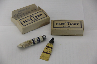
Medicine, The Blue Light Ointment, c1917
... Rub remainder of tube No. 2 Ointment, White, on head and shaft of Penis and Scrotum 5. Hold urine some hours after treatment 6. Report at V.D. ...Rub remainder of tube No. 2 Ointment, White, on head and shaft of Penis and Scrotum 5. Hold urine some hours after treatment 6. Report at V.D. ...Photograph of a box containing two tubes. It was given to Australian soldiers during World War One to prevent venereal disease.Inside box: Directions for Use Before Connection 1. Rub head and shaft of Penis with half the tube of No. 2 Ointment, White. 2. Always wear a sheath After Connection 1. Pass water IMMEDIATELY 2. Wash thoroughly Penis and Scrotum with soap and water. 3. Inject the whole of the contents of the Tube of No. 1. Oitnment, Brown, into pipe and massage back 2 inches. 4. Rub remainder of tube No. 2 Ointment, White, on head and shaft of Penis and Scrotum 5. Hold urine some hours after treatment 6. Report at V.D. Prevention Depot (Blue Light Depot) as soon as possible. 7. Having read directions and understood them, destroy by tearing up or by burning.world war one, medicine, ointment, venereal disease, vd, world war 1, world war, mmm -
 Federation University Historical Collection
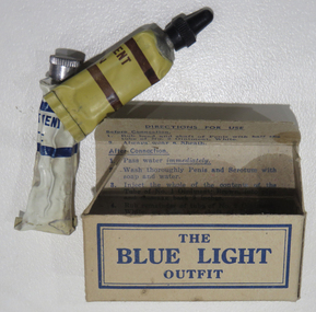
Object, Blue Light Outfit, c1914
... Rub remainder of tube No. 2 Ointment, White, on head and shaft of Penis and Scrotum 5. Hold urine some hours after treatment 6. Report at V.D. ...Rub remainder of tube No. 2 Ointment, White, on head and shaft of Penis and Scrotum 5. Hold urine some hours after treatment 6. Report at V.D. ...The bluelight outfit was offered to members of the armed services in an attempt to prevent venereal disease. Around 60,000 Australian soldiers had contracted venereal disease by the end of the First World War. (http://www.canberratimes.com.au/act-news/secret-wwi-history-of-australian-soldiers-with-venereal-disease-20141022-119wan.html, accessed 21 August 2017) Australia colloquial language defines a Blue Light clinic as a venereal disease clinic.A cardboard box with two tubes of ointment - an anti-Venereal Disease outfit supplied to Australian Armed Services Tube number one is filled with brown contents known as 'No. 1 ointment BROWN' and has a long, tapered opening, with black pastic lid. Tube number two is filled with a thick white ointment 'No. 2 ointment WHITE', with a normal opening and metal lid. The tubes contained 3 percent Argyrol Jelly for gonorrhea and 33% Calomel Ointment (Mercury-Chloride) for syphilis prophylaxis.Inside box: Directions for Use Before Connection 1. Rub head and shaft of Penis with half the tube of No. 2 Ointment, White. 2. Always wear a sheath After Connection 1. Pass water IMMEDIATELY 2. Wash thoroughly Penis and Scrotum with soap and water. 3. Inject the whole of the contents of the Tube of No. 1. Oitnment, Brown, into pipe and massage back 2 inches. 4. Rub remainder of tube No. 2 Ointment, White, on head and shaft of Penis and Scrotum 5. Hold urine some hours after treatment 6. Report at V.D. Prevention Depot (Blue Light Depot) as soon as possible. 7. Having read directions and understood them, destroy by tearing up or by burning.blue light outfit, veneral disease, vd, armed services, gonorrhea, syphilis, disease, medical -
 Northern District School of Nursing. Managed by Bendigo Historical Society Inc.
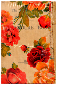
Book - Northern District School of Nursing Graduates Association - Procedure Book for a Student Nurse, 1952 to 1956
... Artificial feeding methods Bandaging, bathing, and bedmaking Care of bedlinen, patient clothing, and valuables Catheterization and care of catheters Charting vital signs and fluid balanceCleaning and sterilizing equipment Counter irritants, enemata, feeding, hygiene, infant care Inhalations, injections, specimen collection, sterilization Special procedures, tray preparation, urine testing, and more Emphasizes the importance of accurate record-keeping, report writing, and communication between staff. ...Artificial feeding methods Bandaging, bathing, and bedmaking Care of bedlinen, patient clothing, and valuables Catheterization and care of catheters Charting vital signs and fluid balanceCleaning and sterilizing equipment Counter irritants, enemata, feeding, hygiene, infant care Inhalations, injections, specimen collection, sterilization Special procedures, tray preparation, urine testing, and more Emphasizes the importance of accurate record-keeping, report writing, and communication between staff. ...The Northern District School of Nursing in Victoria, Australia Graduates Association: History of the School Managed by a committee including hospital administrators, medical professionals, and nursing leaders from several regional hospitals. Associated with major hospitals in the Northern District, including Bendigo Base, Mildura Base, Castlemaine, Echuca, Swan Hill, St. Arnaud, and Kyneton District Hospitals. Provide high-quality theoretical and practical nursing training. Raise the standard of nursing education in the region. Maintain the highest principles of nursing practice. Applicants must be at least 17 years old and hold a Proficiency Certificate or higher educational qualification. Application involves submitting forms, certificates, an interview, an aptitude test, and a medical examination. A three-month probationary period follows initial acceptance. Preliminary Training School: Four-week introductory course covering basic nursing, anatomy, hygiene, and practical skills. Practical experience in various hospital departments, guided by senior staff. Study Block Plan: Includes several blocks of theoretical and practical instruction throughout the three-year program. Regular school exams and two state exams (First Professional and Final State) are required for progression and graduation. Nurses work a 40-hour week with at least one or two days off per week and three weeks of annual leave. Accommodation is provided in comfortable hostels with good facilities. Uniforms are supplied free; nurses provide their own shoes and stockings. Salaries and allowances are regulated, with deductions for board and free medical care. Additional Information Post-Graduate Opportunities. Senior positions require further experience and additional certificates (e.g., Midwifery, Infant Welfare).Diploma courses available in administration, teaching, and specialized nursing fields. Bursaries are available for advanced study.Career Prospects: Graduates can pursue roles in administration, teaching, ward and departmental leadership, district and visiting nursing, industrial and school nursing, and more. Opportunities existed both within hospitals and in community or specialized settings.FAQs and Practical Details. Minimal training costs (mainly exam fees and personal items).Living out allowances and travel expenses are covered. Four training intakes per year; waiting periods are short if qualifications are met.Northern District School of Nursing Graduates Association - Procedure (Report) Book for a Student Nurse 1952 to 1956, that belonged to Ann Rosemary Haigh Archibald 14 pages, also Joan Stot and Jeanette Mary Jobe This item contains the following documents: 3793.2a Procedure Book for a Student Nurse 1952 to 1956, that belonged to Ann Rosemary Haigh Archibald 3793.2b Procedure Book for a Student Nurse 1952 to 1956, that belonged to Joan Stot Front Cover 3793.2c Procedure Book for a Student Nurse 1952 to 1956, that belonged to Jeanette Mary Jobe Front Cover This document is a procedural guide for student nurses, outlining the requirements for recording clinical procedures, charting, and examination preparation. The Procedure Book is filled out by the Sister Tutor in the Training School and by Sisters in wards or departments. Entries in the ward columns are initialed by the supervising Sister.Comments on performance can be added in the Remarks column, which must be initialed and dated. Each student nurse is responsible for carrying their Procedure Book when moving between wards or hospitals. Procedure Books are subject to inspection by Matron, Nursing Supervisor, or Sister Tutor and must be brought to lectures and demonstrations. Marking Code for Procedures D: DemonstratedP: Practised in ClassroomS: Practised under supervision in the wards(Blank): Procedure Books must be presented during the Practical and Oral Sections of the First Professional and State Final Examinations if requested by Examiners. The document contains detailed tables listing required nursing procedures, including: Admission and discharge of patients Preparation and administration of anaesthetics. Artificial feeding methods Bandaging, bathing, and bedmaking Care of bedlinen, patient clothing, and valuables Catheterization and care of catheters Charting vital signs and fluid balanceCleaning and sterilizing equipment Counter irritants, enemata, feeding, hygiene, infant care Inhalations, injections, specimen collection, sterilization Special procedures, tray preparation, urine testing, and more Emphasizes the importance of accurate record-keeping, report writing, and communication between staff. The document includes an index for quick reference to procedures and topics.This guide ensures that student nurses are systematically trained, supervised, and assessed on a wide range of essential clinical skills, with clear documentation and accountability throughout their training.nurse training, lister house, procedure report book, ann archibald, joan stot, jeanette jobe -
 Northern District School of Nursing. Managed by Bendigo Historical Society Inc.
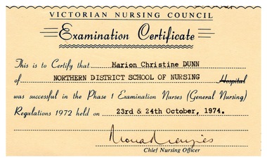
Administrative record - Northern District School of Nursing Progress Report for Nurse M. C. Dunn and other items pertaining to her exams, 1974
... Generally strong answers on patient care, conversions between measurement systems, and infection prevention.Areas for improvement: dilution of lotions, suitable lotions for skin prep, definitions of pH and specific gravity of urine, and details on sterilization (especially destruction of spores). ...Generally strong answers on patient care, conversions between measurement systems, and infection prevention.Areas for improvement: dilution of lotions, suitable lotions for skin prep, definitions of pH and specific gravity of urine, and details on sterilization (especially destruction of spores). ...Managed by a committee including hospital administrators, medical professionals, and nursing leaders from several regional hospitals. Associated with major hospitals in the Northern District, including Bendigo Base, Mildura Base, Castlemaine, Echuca, Swan Hill, St. Arnaud, and Kyneton District Hospitals. Provide high-quality theoretical and practical nursing training. Raise the standard of nursing education in the region. Maintain the highest principles of nursing practice. Applicants must be at least 17 years old and hold a Proficiency Certificate or higher educational qualification. Application involves submitting forms, certificates, an interview, an aptitude test, and a medical examination. A three-month probationary period follows initial acceptance. Preliminary Training School: Four-week introductory course covering basic nursing, anatomy, hygiene, and practical skills. Practical experience in various hospital departments, guided by senior staff. Study Block Plan: Includes several blocks of theoretical and practical instruction throughout the three-year program. Regular school exams and two state exams (First Professional and Final State) are required for progression and graduation. Nurses work a 40-hour week with at least one or two days off per week and three weeks of annual leave. Accommodation is provided in comfortable hostels with good facilities. Uniforms are supplied free; nurses provide their own shoes and stockings. Salaries and allowances are regulated, with deductions for board and free medical care. Additional Information Post-Graduate Opportunities. Senior positions require further experience and additional certificates (e.g., Midwifery, Infant Welfare).Diploma courses available in administration, teaching, and specialized nursing fields. Bursaries are available for advanced study.Career Prospects: Graduates can pursue roles in administration, teaching, ward and departmental leadership, district and visiting nursing, industrial and school nursing, and more. Opportunities exist both within hospitals and in community or specialized settings.FAQs and Practical Details. Minimal training costs (mainly exam fees and personal items).Living out allowances and travel expenses are covered. Four training intakes per year; waiting periods are short if qualifications are met.Northern District School of Nursing Progress Report for Nurse M. C. Dunn and other items pertaining to her exams. This item contains the following six documents: 3791.15a This document is a progress examination report for Nurse M.C. Dunn from the Northern District School of Nursing, summarizing her performance in the Phase 1 Progress Examination held in July 1974. Examination Results Outstanding Performance Passed both Anatomy & Physiology and General Nursing with distinction. Eligible to attend the First Year Study Block and required to sit for the First Professional State Examination in October 1974. Chemistry & Physics will be included in the upcoming examination, with no repeat lectures during the study block. Examiner's General Comments: Anatomy & PhysiologyMost questions were well answered, especially on tissue distribution, limb movement, and muscle functions.Areas needing revision: functions of the skin, diagrams (vertebra, kidney, leaf), function of chordae tendinae, factors maintaining venous return, difference between inspired and expired air, and formation/circulation of cerebrospinal fluid. Some gaps in knowledge about blood volume, urea formation, and water contamination. Generally strong answers on patient care, conversions between measurement systems, and infection prevention.Areas for improvement: dilution of lotions, suitable lotions for skin prep, definitions of pH and specific gravity of urine, and details on sterilization (especially destruction of spores). Good understanding of pressure sore prevention, patient bathing, and care of valuables, though some answers lacked detail or used outdated expressions. 3791.15b This document is a formal acceptance letter addressed to Marion from the Northern District School of Nursing. Marion has been accepted into the Preliminary Training School at the Northern District School of Nursing. Medical and Dental ApprovalHer medical and dental certificates have been reviewed and approved. Marion is scheduled to begin training on 4th February. She is expected to attend on Thursday, 31st January, 1974, prior to the official start. 3791.15c The Victorian Nursing Council's examination instructions for nursing candidates 3791.15d Invitation to a dinner to congratulate the 1973 pilot students in their examination successes 3791.15e Signature of Examiner Card Victorian Nursing Council 3791.15f Examination certificate from the Victorian Nursing Councilnurse training, lister house, marion dunn -
 Flagstaff Hill Maritime Museum and Village
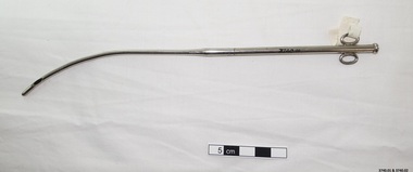
Equipment - Catheter, 20th century
... He believed that chronic stagnant urine residuals and overstretching of the bladder were also responsible. ...He believed that chronic stagnant urine residuals and overstretching of the bladder were also responsible. ...The word “catheter” comes from Greek, meaning “to let or send down.” Catheters were used as early as 3,000 B.C. to relieve painful urinary retention. In those times, many materials were used to form a hollow catheter shape, including straw, rolled up palm leaves, hollow tops of onions, as well as, gold, silver, copper, brass, and lead. Malleable catheters were developed in the 11th century. In time, silver was used as the basis of catheters as it could be bent to any desired shape and was felt to have an antiseptic function. Benjamin Franklin, the inventor and colonial statesman, fashioned silver catheters for use by his older brother John. John suffered from kidney stones and needed to undergo a daily ritual of placing a bulky metal catheter into his bladder. To make these daily requirements on his brother less painful, Franklin worked with his local silversmith on his design for a flexible catheter. "It is as flexible as would be expected in a thing of the kind, and I imagine will readily comply with the turns of the passage," he wrote to John. Holes were bored into the sides of the catheter to allow for drainage. Coudé tip catheters were developed in the 18th and 19th centuries to facilitate male catheterization and continue to be used for this purpose in current medical practice. Catheters made from rubber were developed in the 18th century but were weak at body temperature, leaving debris in the bladder. The advent of rubber vulcanization, by Goodyear in 1844, improved the firmness and durability of the catheter, and allowed for mass production. Latex rubber became available in the 1930s. Dr. Frederic E.B. Foley (a St. Paul urologist) introduced the latex balloon catheter at a urologic meeting in 1935. Though he lost a legal battle with Davol for the patent, this catheter has since been known as the “Foley.” The earliest self-retaining catheters had wing tips (called Malecot) or flexible shoulders (called Pezzer), and were tied to the male penis or sutured to the female labia. Charriere’s French scale was used to describe the external diameter of a catheter. Thus the term “French (Fr)” size was coined. Joseph-Frederic-Benoit Charriere was a 19th century Parisian maker of surgical instruments. A 12 French catheter is approximately 4 mm in external diameter (0.33 mm = 1 French [Fr]). In French-speaking countries, these catheters may be referred to as the Charriere or abbreviated Ch. Catheterization of the bladder was felt to be fairly safe because of the antiseptic principles of Lister (1867). But many physicians continued to be concerned about catheter-related infections as patients were still developing “catheter fever” (systemic infection) despite antiseptic principles. After World War II, Sir Ludwig Guttman introduced the concept of sterile intermittent catheterization in patients with spinal cord injury. For many years, sterile technique was used for catheterization. In 1971, Dr. Jack Lapides of the University of Michigan at Ann Arbor introduced the clean intermittent catheterization (CIC) technique. Dr. Lapides’ theory was that bacteria weren’t the only cause of infection. He believed that chronic stagnant urine residuals and overstretching of the bladder were also responsible. But the fact that CIC was not performed in totally sterile conditions, Dr. Lapides still felt it was superior to indwelling catheters. Initially, Lapides was scorned in the urology world. Three decades after this debate, clean intermittent catheterization remains the preferred method to treat chronic urine retention and neurogenic bladder. Recent regulatory changes have recommended against the reuse of catheters for CIC in an attempt to further reduce the risk of catheter-associated urinary tract infections. https://www.urotoday.com/urinary-catheters-home/history-of-urinary-catheters.html This catheter was donated to Flagstaff Hill Maritime Village by the family of Doctor William Roy Angus, Surgeon and Oculist. It is part of the “W.R. Angus Collection” that includes historical medical equipment, surgical instruments and material once belonging to Dr Edward Ryan and Dr Thomas Francis Ryan, (both of Nhill, Victoria) as well as Dr Angus’ own belongings. The Collection’s history spans the medical practices of the two Doctors Ryan, from 1885-1926 plus that of Dr Angus, up until 1969. ABOUT THE “W.R.ANGUS COLLECTION” Doctor William Roy Angus M.B., B.S., Adel., 1923, F.R.C.S. Edin.,1928 (also known as Dr Roy Angus) was born in Murrumbeena, Victoria in 1901 and lived until 1970. He qualified as a doctor in 1923 at University of Adelaide, was Resident Medical Officer at the Royal Adelaide Hospital in 1924 and for a period was house surgeon to Sir (then Mr.) Henry Simpson Newland. Dr Angus was briefly an Assistant to Dr Riddell of Kapunda, then commenced private practice at Curramulka, Yorke Peninsula, SA, where he was physician, surgeon and chemist. In 1926, he was appointed as new Medical Assistant to Dr Thomas Francis Ryan (T.F. Ryan, or Tom), in Nhill, Victoria, where his experiences included radiology and pharmacy. In 1927 he was Acting House Surgeon in Dr Tom Ryan’s absence. Dr Angus had become engaged to Gladys Forsyth and they decided he further his studies overseas in the UK in 1927. He studied at London University College Hospital and at Edinburgh Royal Infirmary and in 1928, was awarded FRCS (Fellow from the Royal College of Surgeons), Edinburgh. He worked his passage back to Australia as a Ship’s Surgeon on the on the Australian Commonwealth Line’s T.S.S. Largs Bay. Dr Angus married Gladys in 1929, in Ballarat. (They went on to have one son (Graham 1932, born in SA) and two daughters (Helen (died 12/07/1996) and Berenice (Berry), both born at Mira, Nhill According to Berry, her mother Gladys made a lot of their clothes. She was very talented and did some lovely embroidery including lingerie for her trousseau and beautifully handmade baby clothes. Dr Angus was a ‘flying doctor’ for the A.I.M. (Australian Inland Ministry) Aerial Medical Service in 1928. Its first station was in the remote town of Oodnadatta, where Dr Angus was stationed. He was locum tenens there on North-South Railway at 21 Mile Camp. He took up this ‘flying doctor’ position in response to a call from Dr John Flynn; the organisation was later known as the Flying Doctor Service, then the Royal Flying Doctor Service. A lot of his work during this time involved dental surgery also. Between 1928-1932 he was surgeon at the Curramulka Hospital, Yorke Peninsula, South Australia. In 1933 Dr Angus returned to Nhill and purchased a share of the Nelson Street practice and Mira hospital (a 2 bed ward at the Nelson Street Practice) from Dr Les Middleton one of the Middleton Brothers, the current owners of what previously once Dr Tom Ryan’s practice. Dr Tom and his brother had worked as surgeons included eye surgery. Dr Tom Ryan performed many of his operations in the Mira private hospital on his premises. He had been House Surgeon at the Nhill Hospital 1902-1926. Dr Tom Ryan had one of the only two pieces of radiology equipment in Victoria during his practicing years – The Royal Melbourne Hospital had the other one. Over the years Dr Tom Ryan had gradually set up what was effectively a training school for country general-practitioner-surgeons. Each patient was carefully examined, including using the X-ray machine, and any surgery was discussed and planned with Dr Ryan’s assistants several days in advance. Dr Angus gained experience in using the X-ray machine there during his time as assistant to Dr Ryan. When Dr Angus bought into the Nelson Street premises in Nhill he was also appointed as the Nhill Hospital’s Honorary House Surgeon 1933-1938. His practitioner’s plate from his Nhill surgery is now mounted on the doorway to the Port Medical Office at Flagstaff Hill Maritime Village, Warrnambool. When Dr Angus took up practice in the Dr Edward and Dr Tom Ryan’s old premises he obtained their extensive collection of historical medical equipment and materials spanning 1884-1926. A large part of this collection is now on display at the Port Medical Office at Flagstaff Hill Maritime Village in Warrnambool. In 1939 Dr Angus and his family moved to Warrnambool where he purchased “Birchwood,” the 1852 home and medical practice of Dr John Hunter Henderson, at 214 Koroit Street. (This property was sold in1965 to the State Government and is now the site of the Warrnambool Police Station and an ALDI sore is on the land that was once their tennis court). The Angus family was able to afford gardeners, cooks and maids; their home was a popular place for visiting dignitaries to stay whilst visiting Warrnambool. Dr Angus had his own silk worm farm at home in a Mulberry tree. His young daughter used his centrifuge for spinning the silk. Dr Angus was appointed on a part-time basis as Port Medical Officer (Health Officer) in Warrnambool and held this position until the 1940’s when the government no longer required the service of a Port Medical Officer in Warrnambool; he was thus Warrnambool’s last serving Port Medical Officer. (Masters of immigrant ships arriving in port reported incidents of diseases, illness and death and the Port Medical Officer made a decision on whether the ship required Quarantine and for how long, in this way preventing contagious illness from spreading from new immigrants to the residents already in the colony.) Dr Angus was a member of the Australian Medical Association, for 35 years and surgeon at the Warrnambool Base Hospital 1939-1942, He served with the Australian Department of Defence as a Surgeon Captain during WWII 1942-45, in Ballarat, Victoria, and in Bonegilla, N.S.W., completing his service just before the end of the war due to suffering from a heart attack. During his convalescence he carved an intricate and ‘most artistic’ chess set from the material that dentures were made from. He then studied ophthalmology at the Royal Melbourne Eye and Ear Hospital and created cosmetically superior artificial eyes by pioneering using the intrascleral cartilage. Angus received accolades from the Ophthalmological Society of Australasia for this work. He returned to Warrnambool to commence practice as an ophthalmologist, pioneering in artificial eye improvements. He was Honorary Consultant Ophthalmologist to Warrnambool Base Hospital for 31 years. He made monthly visits to Portland as a visiting surgeon, to perform eye surgery. He represented the Victorian South-West subdivision of the Australian Medical Association as its secretary between 1949 and 1956 and as chairman from 1956 to 1958. In 1968 Dr Angus was elected member of Spain’s Barraquer Institute of Barcelona after his research work in Intrasclearal cartilage grafting, becoming one of the few Australian ophthalmologists to receive this honour, and in the following year presented his final paper on Living Intrasclearal Cartilage Implants at the Inaugural Meeting of the Australian College of Ophthalmologists in Melbourne In his personal life Dr Angus was a Presbyterian and treated Sunday as a Sabbath, a day of rest. He would visit 3 or 4 country patients on a Sunday, taking his children along ‘for the ride’ and to visit with him. Sunday evenings he would play the pianola and sing Scottish songs to his family. One of Dr Angus’ patients was Margaret MacKenzie, author of a book on local shipwrecks that she’d seen as an eye witness from the late 1880’s in Peterborough, Victoria. In the early 1950’s Dr Angus, painted a picture of a shipwreck for the cover jacket of Margaret’s book, Shipwrecks and More Shipwrecks. She was blind in later life and her daughter wrote the actual book for her. Dr Angus and his wife Gladys were very involved in Warrnambool’s society with a strong interest in civic affairs. He had an interest in people and the community. They were both involved in the creation of Flagstaff Hill, including the layout of the gardens. After his death (28th March 1970) his family requested his practitioner’s plate, medical instruments and some personal belongings be displayed in the Port Medical Office surgery at Flagstaff Hill Maritime Village, and be called the “W. R. Angus Collection”. The W.R. Angus Collection is significant for still being located at the site it is connected with, Doctor Angus being the last Port Medical Officer in Warrnambool. The collection of medical instruments and other equipment is culturally significant, being an historical example of medicine, administration, household equipment and clothing from late 19th to mid-20th century. Dr Angus assisted Dr Tom Ryan, a pioneer in the use of X-rays and in ocular surgery. Stainless steel catheter with hollow tip from W.R. Angus Collection. Top and end of this instrument screw together. flagstaff hill, warrnambool, shipwrecked coast, flagstaff hill maritime museum, maritime museum, shipwreck coast, flagstaff hill maritime village, great ocean road, dr w r angus, dr ryan, surgical instrument, t.s.s. largs bay, warrnambool base hospital, nhill base hospital, mira hospital, flying doctor, department of defence australia, australian army, army uniform, medical treatment, medical history, medical education, catheter -
 Alfred Hospital Nurses League - Nursing History Collection
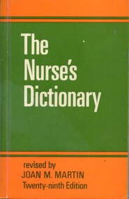
Book - Illustrated dictionary, Honnor Morten 1861-1913, The Nurse's Dictionary, 1980
... Over the years the dictionary has been updated by various editors to keep pace with evolving nursing practices and nedical terminology Insight into nursing knowledge and practice in 1980 nursing-dictionary medical-dictionary Provides definitions for medical and nursing terminology includes appendices on first aid, biochemistry, urine testing and diet on front end pag at top right 'W I THOMAS [blue ink]/Reference [black ink] Illustrated book. ...Provides definitions for medical and nursing terminology includes appendices on first aid, biochemistry, urine testing and dietIllustrated book. On front cover the title [white ink] is printed on green square. Orange bannd above and below, on lower band revisor and edition are printed [black ink] Same information brinted on spine.Book has been covered with clear adhesive filmnon-fictionProvides definitions for medical and nursing terminology includes appendices on first aid, biochemistry, urine testing and dietnursing-dictionary, medical-dictionary