Showing 176 items matching "obstetric"
-
 Royal Australian and New Zealand College of Obstetricians & Gynaecologists (RANZCOG)
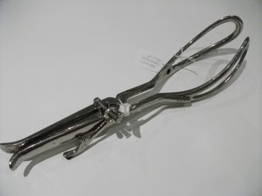
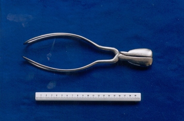
Forceps, Tarnier's
Tarnier's forceps, metal with one traction arm, second arm missing. Stamped "5" on both inner forcep blades.obstetric delivery -
 Royal Australian and New Zealand College of Obstetricians & Gynaecologists (RANZCOG)
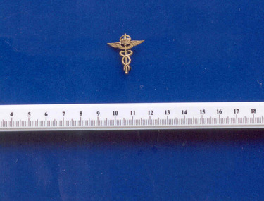
Badge - Royal Air Force (RAF) Medical Branch collar insignia worn by F J Browne, World War I, Firmin, London
This badge belonged to F.J. Browne and would have been worn during World War I. Hermes (Mercury) was the messenger of the gods and known for carrying a staff known as the Caduceus. The caduceus included two snakes topped off with a set of wings. Caduceus is from the Greek root meaning “herald’s wand”.Francis James Browne died in Sydney 1963. He had a long career in obstetrics and gynaecology. Summary of appointments include: General Practice in Wales, Maternity Department of the Edinburgh Royal Infirmary, 1st director of obstetric unit, University College Hospital London. Retired and continued postgraduate teaching in London and NSW. Married to Grace Cuthbert, who was director of Maternal and Baby Welfare in NSW. Collection of objects transferred from the Archives to the Museum collection found amongst Professor FJ Browne's papers.Brass badge depicting a crown mounted on top of a caduceus (image of two snakes wrapped around a staff topped by wings). Clip attached to back of badge is inscribed 'FIRMIN LONDON'.numismatics, browne fj, world war i -
 Royal Australian and New Zealand College of Obstetricians & Gynaecologists (RANZCOG)
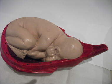
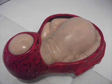
Obstetric teaching model
Originally this six part teaching model was used and owned by the former Queen Victoria Womens' Hospital, Lonsdale/ Swanston Streets, Melbourne. With the amalgamation of hospitals, the models were transferred to the Monash Medical Centre, Clayton.Foetus in uterus, cervix not dilated. Painted plaster. The birth process in six stages; first of six models.teaching aid, obstetric delivery -
 Royal Australian and New Zealand College of Obstetricians & Gynaecologists (RANZCOG)
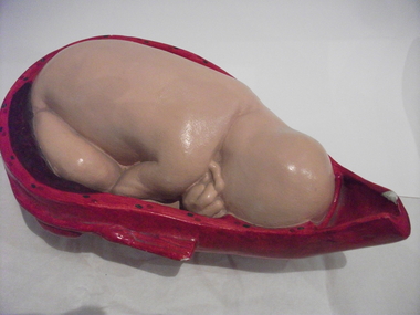
Obstetric teaching model
Originally this six part teaching model was used and owned by the former Queen Victoria Womens' Hospital, Lonsdale/ Swanston Streets, Melbourne. With the amalgamation of hospitals, the models were transferred to the Monash Medical Centre, Clayton.Foetus in uterus, head engaged, cervix partially dilated.. Painted plaster. The birth process in six stages;second of six models.teaching aid, obstetric delivery -
 Royal Australian and New Zealand College of Obstetricians & Gynaecologists (RANZCOG)
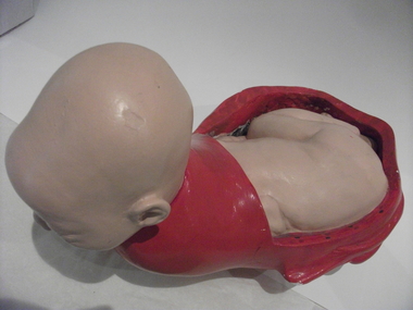
Obstetric teaching model
Originally this six part teaching model was used and owned by the former Queen Victoria Womens' Hospital, Lonsdale/ Swanston Streets, Melbourne. With the amalgamation of hospitals, the models were transferred to the Monash Medical Centre, Clayton.Foetus in uterus, head crowning.. Painted plaster. The birth process in six stages; third of six models.teaching aid, obstetric delivery -
 Royal Australian and New Zealand College of Obstetricians & Gynaecologists (RANZCOG)
Obstetric teaching model
Originally this six part teaching model was used and owned by the former Queen Victoria Womens' Hospital, Lonsdale/ Swanston Streets, Melbourne. With the amalgamation of hospitals, the models were transferred to the Monash Medical Centre, Clayton.Foetus, head fully emerged. Painted plaster. The birth process in six stages; fourth of six models.teaching aid, obstetric delivery -
 Royal Australian and New Zealand College of Obstetricians & Gynaecologists (RANZCOG)
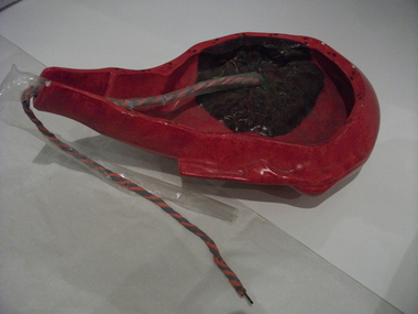
Obstetric teaching model
Originally this six part teaching model was used and owned by the former Queen Victoria Womens' Hospital, Lonsdale/ Swanston Streets, Melbourne. With the amalgamation of hospitals, the models were transferred to the Monash Medical Centre, Clayton.Placenta with attached umbilical cord, uterus still enlarged. Painted plaster. The birth process in six stages; fifth of six models.teaching aid, obstetric delivery -
 Royal Australian and New Zealand College of Obstetricians & Gynaecologists (RANZCOG)
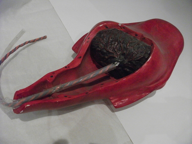
Obstetric teaching model
Originally this six part teaching model was used and owned by the former Queen Victoria Womens' Hospital, Lonsdale/ Swanston Streets, Melbourne. With the amalgamation of hospitals, the models were transferred to the Monash Medical Centre, Clayton.Placenta with attached umbilical cord, uterus smaller, cervix more contracted. [end of series]. Painted plaster. The birth process in six stages;sixth of six models.teaching aid, obstetric delivery -
 Royal Australian and New Zealand College of Obstetricians & Gynaecologists (RANZCOG)
Identification bracelet worn by F J Browne
This bracelet is possibly associated with FJ Browne's service with the Royal Army Medical Corps in World War I. Identification bracelets were worn during World War I and II in England.Francis James Browne died in Sydney 1963. He had a long career in obstetrics and gynaecology. Summary of appointments include: General Practice in Wales, Maternity Department of the Edinburgh Royal Infirmary, 1st director of obstetric unit, University College Hospital London. Retired and continued postgraduate teaching in London and NSW. Married to Grace Cuthbert, who was director of Maternal and Baby Welfare in NSW. Collection of objects transferred from the Archives to the Museum collection found amongst Professor FJ Browne's papers.Silver identification bracelet. Bracelet is engraved "FJ BROWNE/ HEATH LODGE/ WATFORD HEATH." Reverse of bracelet is engraved "SILVER". browne fj -
 Royal Australian and New Zealand College of Obstetricians & Gynaecologists (RANZCOG)
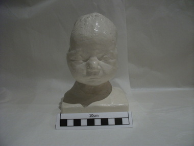
Plaster cast of the head of the first baby to be delivered under anaesthesia by the use of ether, 1847
This a cast of the first baby to be delivered under anaesthesia, by the use of ether, in Edinburgh on 19 January 1847. The famous physician James Young Simpson, Professor of midwifery at Edinburgh University, attended this birth and wrote about it in the Monthly Journal of Medical Science 1846-7 Vol.7, p649-640. The cast of the baby's head was given to Lance Townsend, Professor of Obstetrics and Gynaecology, University of Melbourne by Robert Kellar, then Professor of Midwifery and Diseases of Women at the University of Edinburgh, when Professor Townsend was visiting Edinburgh. There is at least one other plaster copy; one is located at Wood Library-Museum of Anesthesiology, 520 North Northwest Highway, Park Ridge, IL 60068-2573, USAReplica of a new born baby's head, painted plaster, life size. The model of the head shows a large indentation of two and a half inches in the skull on the left side. The baby was delivered through a severely deformed pelvis, suffered a large indentation to the skull and did not live.obstetric delivery, anaesthesia -
 Royal Australian and New Zealand College of Obstetricians & Gynaecologists (RANZCOG)
Tool - Short handled Simpson-type obstetrical forceps, Down, London
First developed by James Young Simpson in 1848, Simpson forceps have become arguably the most popular model of forceps for use, and were adapted in the creation of many later designs.Short handled set of forceps, possibly made of stainless steel. Marked "DOWN LONDON" on inner aspect of left blade handle.'DOWN LONDON'obstetric delivery -
 Royal Australian and New Zealand College of Obstetricians & Gynaecologists (RANZCOG)
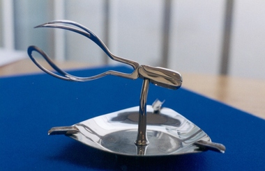
Commemorative ashtray presented to Dr Frank Forster, c. 1965, Rocket & Co, London
This item was a gift to Dr Frank Forster from colleagues (and possibly students) whilst he was working at the Hosptial for Women at Soho, London, c 1965. [Source: Librarian Hilary Belton] The ashtray was made by Rocket & Co, a manufacturer of surgical instruments. Geofrey Bishop and Bryan Hibbard also have similar ashtrays.Chrome ashtray, consisting of a triangular base with three cigarette rests. At the centre of the base, on a central stem/pedestal, is a set of replica miniature forceps.dr frank forster, obstetric delivery, ephemera -
 Royal Australian and New Zealand College of Obstetricians & Gynaecologists (RANZCOG)
Doctor's theatre gown worn by Dr Mitchell Henry O'Sullivan, c. 1930s
The wearing of gown became mandatory in all operating theatres from the 1900s and in 1914-1918 during the Spanish flu epidemic. During the 1930s gowns were worn when attending polio patients. From 1945 onwards, midwifery hospitals required all staff working in labour wards, premature nurseries, and special care (observation nurseries) to wear gowns when in contact with mothers or babies. During the 1950s the gown regime helped to combat the spread of golden stph in midwifery hospitals. Dr Mitchell Henry O'Sullivan worked in the Victorian country town of Casterton as a general practitioner from 1919 until his death in 1977. He also practiced obstetrics. His son, Dr David More O'Sullivan donated his obstetric bag and its contents to the College in 1999. The bag and contents are a unique time capsule of the type of instruments and pharmaceuticals used in the inter-war period.Cotton gown with high round collar and long sleeves. Gown is made in two sections with a centre doubled seam. The collar is made to button at the neck, but the button on this gown is missing. Wrists of gown are fastened with flat mother of pearl buttons. Open at back with six ties. Laundry tag taped to right side of gown.surgery -
 Royal Australian and New Zealand College of Obstetricians & Gynaecologists (RANZCOG)
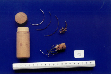
Tool - Collection of suture needles used by Dr Mitchell Henry O'Sullivan
Needles in this collection include: -2 x abdominal triangular cutting edge suture needles, used for skin suture. Size 2/0 -Morrison's half circle round bodied suture needle, size 3 and size 2 -Bonney's regular curved cutting edge suture needle, size 7 or 8 -Regular curved triangular pointed suture needle, size 2 and size 6 -Ferguson's round point half circle suture needle, size 15 -Hagerdorn's reversed 1/20 suture needle, size 7 -Bonney's curved suture needle, size 7 and size 9 -Regular curved triangular cutting point suture needle, size 15 -Boston/intestinal fine round bodied half circle suture needle Dr Mitchell Henry O'Sullivan worked in the Victorian country town of Casterton as a general practitioner from 1919 until his death in 1977. He also practiced obstetrics. His son, Dr David More O'Sullivan donated his obstetric bag and its contents to the College in 1999. The bag and contents are a unique time capsule of the type of instruments and pharmaceuticals used in the inter-war period.Collection of suture needles in wooden case. Consists of four, loose, crescent shaped needles, and an additional nine needles stuck into a piece of cork. Case is cylindrical, has a lid and is possibly made from pine wood.surgery -
 Royal Australian and New Zealand College of Obstetricians & Gynaecologists (RANZCOG)
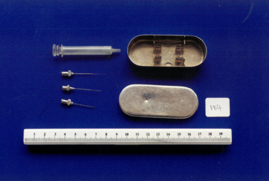
Equipment - Hypodermic syringe and needles used by Dr Mitchell Henry O'Sullivan
Dr Mitchell Henry O'Sullivan worked in the Victorian country town of Casterton as a general practitioner from 1919 until his death in 1977. He also practiced obstetrics. His son, Dr David More O'Sullivan donated his obstetric bag and its contents to the College in 1999. The bag and contents are a unique time capsule of the type of instruments and pharmaceuticals used in the inter-war period.Hypodermic glass syringe (.1) with three hypodermic needles (.2 - .4) and metal storage case (.5 - .6). Barrel of syringe is marked from with measurements from 0-20. .2 is a needle head with a rounded flange tapering towards the needle shaft. .3 and .4 are needle heads with flattened oval bulbs, attached to a round bead which holds the needle shaft. The case is oval in shape with the word 'Crystal' engraved diagonally into the top of the lid. There are two fixings attached inside the bottom of the case which form a cradle for the storage of the syringe.'Crystal' -
 Royal Australian and New Zealand College of Obstetricians & Gynaecologists (RANZCOG)
Tool - Obstetric forceps used by Dr Michael Kloss, Jetter and Scheerer
Jetter and Scheerer were a surgical instrument maker founded in Germany in 1867. Their company symbol is that of a serpent curled around a rod, surmounted by a coronet/crown. This instrument was part of a collection of instruments used by Dr Michael Kloss in his medical practice. Dr Kloss subsequently donated this collection to the College.Metal forceps, consisting of two blades which lock together with a pin fitting. The handle of one of the forceps blades is engraved with the word 'Kloss', both on the outside and on the inner aspect. The number '26' is engraved on the inner aspect of both blades near the pin joint. The upper shaft of one blade is engraved with the word 'HOSON'. The blade is also engraved with a derivation of the Rod of Asclepius, featuring a serpent wrapped around a rod, with the entire design topped by a crown.'Kloss' '26' 'HOSON'obstetrics -
 Royal Australian and New Zealand College of Obstetricians & Gynaecologists (RANZCOG)
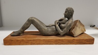
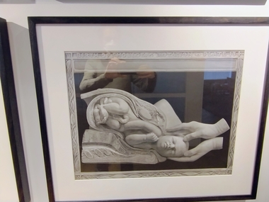
Decorative object - Statue of a mother reclining with a newborn bavy, Victoria Chancellor (nee Simcock)
This sculpture was a commissioned artwork made by Victora Chancellor (nee Simcock), a Sydney artist.Ceramic sculpture, depicting a reclining mother holder her newborn baby. The mother is lying on her back, with knees drawn up and feel flat on the ground, protectively cradling a newborn baby on her chest. The umbilical cord between baby and mother is still attached. The mothers upper back is resting against a rock, holding her partially upright. Sculpture is mounted on a rectangular wooden base.obstetric delivery -
St Vincent's Hospital Melbourne Archives
Certificate - Women's Hospital Midwifery Department Obstetric Nurse certificate awarded to Agnes Mclean
agnes mclean, women's hospital melbourne -
 Royal Australian and New Zealand College of Obstetricians & Gynaecologists (RANZCOG)
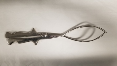
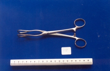
Tool - Guy's tongue forceps used by Dr Mitchell Henry O'Sullivan, 1930 (approximate)
During surgery under general anaesthetic, these forceps were used to pull the tongue forward to keep the patient's airways clear from obstruction. This tool was in general use from the 1930s onwards in teaching hospitals, and became a standard piece of equipment on all anaesthetic trolleys. In the majority of teaching hospitals, the blades of these forceps were smooth and wide to cause less trauma to the tongue. This particular style of tongue forceps is known as Guy's tongue forceps.Dr Mitchell Henry O'Sullivan worked in the Victorian country town of Casterton as a general practitioner from 1919 until his death in 1977. He also practiced obstetrics. His son, Dr David More O'Sullivan donated his obstetric bag and its contents to the College in 1999. The bag and contents are a unique time capsule of the type of instruments and pharmaceuticals used in the inter-war period.Pair of metal forceps. Similar in appearance to a pair of scissors, and other surgical forceps, but with rounded teardrop shaped tips. There is also a notch clip for the handle to keep the forceps open. Inner aspect of both forceps blades engraved with the number '3'. The left hand blade is also engraved with a 'P'.anaesthesia -
 Royal Australian and New Zealand College of Obstetricians & Gynaecologists (RANZCOG)
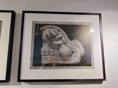
Print - Reproduction print of plate from 'Birth atlas', Maternity Center Association, 1943, Robert Latou Dickinson et al, Plate 10. Full dilation cervix high head deep in pelvis, 1943
A series of six prints showing various stages of labour, donated to the College in 2000.Black and white reproduction print of a plate from a book, enclosed in a wooden frame. Text at the top of the print reads 'FULL DILATION CERVIX HIGH HEAD DEEP IN PELVIS PLATE 10'. The plate depicts a detailed cross section image of a baby in the womb. Label attached to back of work reads: 'Robert Latou DICKINSON and Abram BELSKI/ Plate 10. Full dilation cervix high head deep in pelvis/From: Birth atlas: reproduction of twenty-four life size sculptures of fertilization, growth,/stages of labour and involution./New York: Maternity Center Association, 1943. 6th ed./Gift of St Georges Hospital, Kew, 2000'.FULL DILATION CERVIX HIGH HEAD DEEP IN PELVIS PLATE 10obstetric delivery -
 Royal Australian and New Zealand College of Obstetricians & Gynaecologists (RANZCOG)
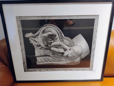
Print - Reproduction print of plate from 'Birth atlas', Maternity Center Association, 1943, Robert Latou Dickinson et al, Plate 11. Abdominal muscle drive pelvic floor thin, 1943
A series of six prints showing various stages of labour, donated to the College in 2000.Black and white reproduction print of a plate from a book, enclosed in a wooden frame. Text at the top of the print reads 'ABDOMINAL MUSCLE DRIVE PELVIC FLOOR THIN PLATE 11'. The plate depicts a detailed cross section image of a baby in the womb. Label attached to back of work reads: 'Robert Latou DICKINSON and Abram BELSKI/ Plate 11. Abdominal muscle drive pelvic floor thin/From: Birth atlas: reproduction of twenty-four life size sculptures of fertilization, growth,/stages of labour and involution./New York: Maternity Center Association, 1943. 6th ed./Gift of St Georges Hospital, Kew, 2000'.ABDOMINAL MUSCLE DRIVE PELVIC FLOOR THIN PLATE 11obstetric delivery -
 Royal Australian and New Zealand College of Obstetricians & Gynaecologists (RANZCOG)
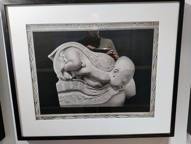
Print - Reproduction print of plate from 'Birth atlas', Maternity Center Association, 1943, Robert Latou Dickinson et al, Plate 12. Head turns upward pelvic floor retreats, 1943
A series of six prints showing various stages of labour, donated to the College in 2000.Black and white reproduction print of a plate from a book, enclosed in a wooden frame. Text at the top of the print reads 'HEAD TURNS UPWARD PELVIC FLOOR RETREATS PLATE 12'. The plate depicts a detailed cross section image of a baby in the womb. Label attached to back of work reads: 'Robert Latou DICKINSON and Abram BELSKI/ Plate 12. Head turns upward pelvic floor retreats/From: Birth atlas: reproduction of twenty-four life size sculptures of fertilization, growth,/stages of labour and involution./New York: Maternity Center Association, 1943. 6th ed./Gift of St Georges Hospital, Kew, 2000'.HEAD TURNS UPWARD PELVIC FLOOR RETREATS PLATE 12obstetric delivery -
 Royal Australian and New Zealand College of Obstetricians & Gynaecologists (RANZCOG)
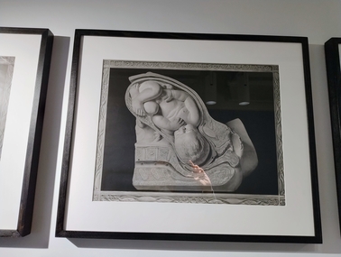
Print - Reproduction print of plate from 'Birth atlas', Maternity Center Association, 1943, Robert Latou Dickinson et al, Plate 9. Labor cervix dilating & bag of waters, 1943
A series of six prints showing various stages of labour, donated to the College in 2000.Black and white reproduction print of a plate from a book, enclosed in a wooden frame. Text at the top of the print reads 'LABOR CERVIX DILATING & BAG OF WATERS PLATE 9'. The plate depicts a detailed cross section image of a baby in the womb. Label attached to back of work reads: 'Robert Latou DICKINSON and Abram BELSKI/ Plate 9. Labor cervix dilating & bag of waters/From: Birth atlas: reproduction of twenty-four life size sculptures of fertilization, growth,/stages of labour and involution./New York: Maternity Center Association, 1943. 6th ed./Gift of St Georges Hospital, Kew, 2000'.LABOR CERVIX DILATING & BAG OF WATERS PLATE 9obstetric delivery -
 Royal Australian and New Zealand College of Obstetricians & Gynaecologists (RANZCOG)
Print - Reproduction print of plate from 'Birth atlas', Maternity Center Association, 1943, Robert Latou Dickinson et al, Plate 13. Birth of shoulders rotation, 1943
A series of six prints showing various stages of labour, donated to the College in 2000.Black and white reproduction print of a plate from a book, enclosed in a wooden frame. Text at the top of the print reads 'BIRTH OF SHOULDERS ROTATION PLATE 13'. The plate depicts a detailed cross section image of a baby in the womb. Label attached to back of work reads: 'Robert Latou DICKINSON and Abram BELSKI/ Plate 13. Birth of shoulders rotation/From: Birth atlas: reproduction of twenty-four life size sculptures of fertilization, growth,/stages of labour and involution./New York: Maternity Center Association, 1943. 6th ed./Gift of St Georges Hospital, Kew, 2000'.BIRTH OF SHOULDERS ROTATION PLATE 13obstetric delivery -
 Royal Australian and New Zealand College of Obstetricians & Gynaecologists (RANZCOG)
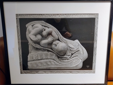
Print - Reproduction print of plate from 'Birth atlas', Maternity Center Association, 1943, Robert Latou Dickinson et al, Plate 8. Before labour, 1943
A series of six prints showing various stages of labour, donated to the College in 2000.Black and white reproduction print of a plate from a book, enclosed in a wooden frame. Text at the top of the print reads 'BEFORE LABOR | BIRTH ATLAS | PLATE 8'. The plate depicts a detailed cross section image of a baby in the womb. Label attached to back of work reads: 'Robert Latou DICKINSON and Abram BELSKI/ Plate 8. Before labour/From: Birth atlas: reproduction of twenty-four life size sculptures of fertilization, growth,/stages of labour and involution./New York: Maternity Center Association, 1943. 6th ed./Gift of St Georges Hospital, Kew, 2000'.BEFORE LABOR | BIRTH ATLAS | PLATE 8obstetric delivery -
 Royal Australian and New Zealand College of Obstetricians & Gynaecologists (RANZCOG)
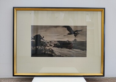
Print - Lithograph, William Balfour Ker (1877-1918), A Hurry Call, The Doctor v The Stork, 1905
William Balfour Ker was a Canadian-American artist. He was also a declared socialist, and his political stance was often reflected in his art. This lithograph is a compelling illustration of the race against time sometimes faced by doctors when trying to get to a patient to assist in birth before the baby is born.A black and white lithograph. At top right, a stork is flying through the air, with a baby bundled in a cloth that it is holding in its beak. To the left, a horse drawn buggy follows rapidly behind, with the driver wielding a whip and urging on a speeding white horse. The artist's signature printed at bottom right corner of image reads 'WM.BALFOUR-KER'. The lithograph has been mounted and framed in wooden frame. The outer edge of the frame is black, and the inner edge is gold. On the back of the object, a small piece of paper bearing the title of the lithograph has been attached at bottom centre. It reads 'A HURRY CALL, THE DOCTOR v THE STORK'. A second tag attached to the back of the work reads 'From FF', suggesting the donor of this object was Frank Forster. An old display label is attached to the bottom left of the back of the work. A wire and two hooks have been attached to the back of the frame for hanging.obstetric delivery